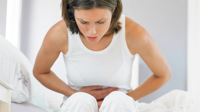
Examination by a doctor
Depending on the stage of development of the disease, dry skin and tongue may occur due to dehydration. When a doctor palpates the abdominal area, the patient may feel sharp pain, sometimes reaching peritonitis, which is tension in the abdominal region due to inflammation. This indicates the appearance of holes in the walls of the large intestine. A good result is obtained by a digital examination of the rectum, which allows you to find all purulent formations in the wall of the rectum, fistulas, cracks, compactions or tuberosity of the rectum. This method will help detect the presence of blood, pus and mucus.
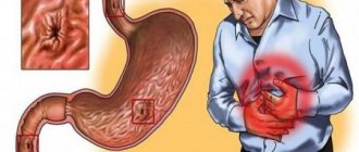
Main manifestations of the disease:
- Frequent loose stools. Bowel movements occur up to 20 times a day, in severe cases - up to 40. Fecal incontinence may occur.
- Impurities of blood, mucus, pus in the stool.
- Abdominal pain of a cramping nature. Most often they occur in the lower left abdomen, sometimes near the navel. They intensify before defecation and disappear after it.
Many patients have a decrease in appetite, they refuse food, as pain and diarrhea intensify after eating. Weight decreases, a person loses weight, and constantly experiences weakness.
Take care of yourself, book a consultation now
Analyzes
For ulcerative colitis, laboratory diagnosis involves examining a patient who will have to undergo:
- General blood analysis. Such a standard analysis will allow us to find an inflammatory process, which is characterized by an increase in the level of leukocytes in the blood. This figure is 9.0 * 10 to the 9th power / l. An increase in ESR is also observed, but with internal bleeding, the hemoglobin level, on the contrary, decreases, depending on the amount of blood lost.
- Stool analysis. The examination can determine the presence of hidden blood in the stool; Blood chemistry. An increase in C-reactive protein in the body and a decrease in total protein indicate inflammation. According to statistics, antineutrophil cytoplasmic antibodies are detected in 60% of patients. These microorganisms act against neutrophils, which tend to attack and cause inflammation.
- Microbiological research. The patient must submit cultures taken from the mucous membrane to rule out an infectious form of the disease. The body is checked for the presence of pathogenic flora, for different types of viruses, chlamydia, and helminths.
Colonoscopy or endoscopic examination of the colon
The procedure is carried out with histological examination and biopsy. This method will accurately determine a nonspecific disease. A study at the cellular level is carried out to make a more accurate diagnosis, because there are similarities between ulcerative colitis and Crohn's disease due to a similar endoscopic picture. During colonoscopy, redness and swelling of the sigmoid and colon mucosa, as well as other areas of the large intestine, appear. The study smoothes out rib-like protrusions in the rectum, making it corrugated.
Endoscopy helps to detect ulcerative formations of different sizes in the mucous membrane, depending on the stage of the lesion, or areas with bleeding. Such defects are often found with moderate and severe severity of the disease. The ulcers vary in size, and their bottom is sometimes covered with fibrin or purulent discharge. In the middle stage of ulcerative colitis, ulcerative formations may be absent, although the mucous membrane will be marked with a pattern in the form of small grains. If the study showed the presence of only one ulcer, then this may indicate intestinal cancer, although there is no need to panic ahead of time. In this case, the doctor takes a piece of tissue from the detected defect for biopsy and further diagnosis. A colonoscopy will help determine the severity and duration of the disease.
Symptoms
Manifestations of the disease depend on the form of its course and on how large a section of the intestine is involved in the process. Nonspecific ulcerative colitis can occur in one of three forms:
- The acute form is rare. Usually the process involves the entire intestine, symptoms increase quickly, the disease is severe, and complications develop early.
- In the chronic form, symptoms increase gradually and persist permanently.
- The most common form is recurrent. An exacerbation occurs, then the symptoms subside and disappear for 3–6 months. After this, a new exacerbation occurs.
Magnetic resonance imaging
MRI machine
MRI allows you to check any diseased organ at the cellular level. The diagnostic method is based on the ability of atomic nuclei to respond to electromagnetic waves. Often, contrast is used during the procedure for a better diagnosis of ulcer. The liquid consists of a fat emulsion with gadolinium or iron oxides. To reduce intestinal motility, antispasmodics are used, which are administered intravenously or added to drinking water. This helps to improve the procedure for examining the mucous membrane.
For a more effective examination, artificial intestinal distension is used using a rectal enema or oral administration of contrast. To reduce the absorption of the drug into the patient’s body, special compounds are used to slow down this process. This diagnostic method is not recommended for people suffering from claustrophobia, epileptic and convulsive seizures. Before the procedure, the doctor must be aware of the presence of allergic reactions to the solution used, patches on the body or tattoos.
X-ray
This type of diagnosis determines toxic megacolon. This is an enlargement of the colon that can be life-threatening. X-ray examination helps to detect the accumulation of gases in the transverse intestine or in the abdominal cavity, which indicates perforation of a peptic ulcer. The disappearance of haustra and unevenness of the pattern due to the development of ulcerative formations are noted. With a prolonged, severe illness, the lumen of the colon is significantly narrowed, and the intestinal walls are characterized by increased rigidity. Exacerbation of the disease shortens the colon due to the inflammatory process in the human body.
Pathomorphological diagnosis
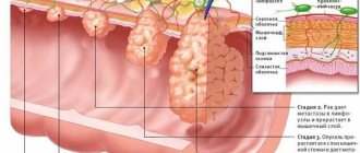
The study can reveal what stage of development a nonspecific illness has due to the symptoms, examination and complaints of the patient:
- The mild stage involves swelling and inflammation of the mucous membrane.
- The average degree is marked by inflammation, the development of ulcerative defects and slight bleeding.
- A severe form of the disease is the multiple appearance of ulcerative formations, which lead to smoothing and loss of relief of the mucous membrane. Due to the active recovery process, pseudopolyps appear. Often this phase is due to an increased risk of developing toxicosis, which causes inflammation of the abdominal area and bloating. With such symptoms, another type of diagnosis is contraindicated.
Treatment with medications
Effective treatment of ulcerative colitis is possible only with a qualified doctor. In case of acute ulcerative colitis, the patient is in the hospital, where he observes strict bed rest until the intensity of the symptoms decreases. At the time of remission, the person continues to lead a normal lifestyle, taking into account the recommendations of the attending physician regarding medication and diet.
Drug therapy for colitis includes:
In some cases, the doctor prescribes Cyclosporin-A, which is relevant for the rapid development of UC in the acute phase. A dose of 4 mg per 1 kg of human weight is administered intravenously. Symptomatic treatment of nonspecific colitis involves taking painkillers (Ibuprofen, Paracetamol and others) and vitamins B, C.
UC in a child can be cured by following a diet. Doctors in 95% prescribe “dairy-free table No. 4 according to Pevzner.” The menu mainly consists of protein through the consumption of meat, fish and eggs.
The basis of drug treatment for colitis in children is Sulfasalazine and other medications that contain Mesalazine. The drugs are taken orally or administered through enemas or suppositories. Dosage and course are determined on a strictly individual basis. Along with these measures, symptoms are eliminated.
However, if adequate therapy is not available, then there is a risk of developing complications of colitis, which occur as follows:
If you do not start treating the disease in a timely manner, then in 7-10% of cases this leads to death, and in 45-50% - to disability.
Irrigoscopy
Picture after irrigoscopy
This type of study is carried out to obtain complete information about the condition of the large intestine, its relief and size. Diagnosis of ulcerative colitis is less traumatic, therefore it is recommended for patients who cannot undergo colonoscopy for any reason. After the procedure, the doctor will know about the condition of the intestinal walls not only in straight sections, but also in bends.
Using an enema, a contrast agent is administered after preliminary cleansing of the intestines. Then several pictures are taken as the patient's body position changes. After this, the colon is emptied of contrast, allowing its ability to contract and its relief to be studied. If clearer X-rays are required, the intestines are filled with air. This diagnostic method is called double contrast. The substance remaining on the walls of the intestine helps to examine its back wall in more detail.
This study is not used in people with long intestines and in weakened patients. The procedure is also prohibited if intestinal obstruction is suspected. Irrigoscopy involves the use of water-soluble contrast when there is a risk of perforation of the intestinal walls.
Why does ulcerative colitis occur?
Modern scientists cannot accurately name the reasons that cause UC. There are known risk factors that contribute to the development of the disease:
- Impaired immune function. Some scientists believe that a viral or bacterial infection can cause the immune system to malfunction and attack the intestinal lining. According to another point of view, infection has nothing to do with it, and UC is an autoimmune condition associated with defects in the immune system.
- Heredity. Recent research has identified genes that scientists believe contribute to the development of ulcerative colitis. However, the role of heredity is not very large - only 20% of patients have sick relatives.
- Environmental pollution. According to statistics from the United States and Western European countries, the disease is more common in people who live in large cities.
- Cigarette smoke and unfavorable sanitary and hygienic living conditions.
- High content of linoleic acid in the diet. It is present in red meat and margarine. In the course of research, scientists found that a third of patients with ulcerative colitis consume large amounts of linoleic acid.
Sigmoidoscopy
Differential diagnosis is carried out using a proctoscope to examine the rectum and all parts of the sigmoid colon. The device for the procedure is a rigid tube, the length of which reaches 30 cm, and the diameter is 2 cm. The device is equipped with a special apparatus for supplying air, a light source and lenses. Thanks to the study, the doctor is able to see the condition of the mucous membrane and find such neoplasms as cracks, tumors, polyps, hemorrhoids, scars, etc. If the need arises, a biopsy can be performed.
Sigmoidoscopy does not take much time and is performed in the hospital. The patient has to remove all clothing below the waist and take a knee-elbow position or lie on his side. First, the doctor examines the rectum with his fingers, after which he inserts a rectoscope 5 cm into the anus. The remaining manipulations are carried out through visual observation, when the device moves only along the intestinal canal.
Ultrasound
Ultrasound examination helps to quickly and effectively determine the location, size and condition of the intestine in ulcerative colitis. This research method is completely safe, allowing it to be used in almost all areas of medicine. Basically, the procedure is performed if the development of a disease in the abdominal part of the body is suspected. Doctors advise doing an ultrasound of the intestines to determine the thickness of the walls of the organ being examined, preventing the occurrence of various ailments. Such a study is indispensable for dynamic monitoring of patients with ulcerative colitis, determining the effectiveness of the prescribed therapy.
Prevention
To avoid the development of erosive colitis, you must:
- undergo a general medical examination at least once a year;
- forget about bad habits;
- eat comprehensively and properly;
- to live an active lifestyle;
- strengthen the immune system.
Erosive colitis is an insidious disease that greatly disrupts a person’s normal life activities. At the first symptoms, you should consult a doctor, because it can cause serious complications.
Colitis of the intestine Erosive gastritis Ulcerative colitis of the intestine Spastic colitis Irritable bowel syndrome Bowel cancer
CT scan
CT scanning is often called virtual colonoscopy. To obtain a complete picture of the inside of the large intestine, minimal doses of X-ray radiation are used during the procedure. The examination takes no more than 20 minutes and is completely painless. This diagnostic method will help identify thickening of the colon wall and nonspecific colitis.
During the procedure, the patient lies down on a special table, and a tube is inserted into the anus to a depth of 5 cm to supply air, allowing the colon to straighten. Then the patient is placed together with the table in the X-ray machine. During operation, the device begins to rotate in a spiral, taking pictures from different angles. For a more qualitative study, an iodine-based contrast solution is used. The liquid is administered by rectal enema. It does not have the ability to be absorbed into the intestines, and only the mucous membrane of the organ can be stained.