Benign tumors of the duodenum are rare.
Thus, according to the Mayo Clinic (USA), out of 6044 cases of tumor lesions of this organ, only 44 cases had benign neoplasms.
They account for an average of 20.3% of all benign tumors of the small intestine.
According to A. Essinger (1963), during autopsy they are found in 0.002% of cases.
Benign tumors of the duodenum are very diverse in their origin and morphological characteristics. The most common are adenomas and adenomatous polyps, as well as leiomyomas and lipomas, accounting for 13 to 19.8%.
We observed 36 patients with benign tumors of the duodenum. Adenomatous polyps were present in 22 patients, leiomyomas in 6, neurilemmomas in 5, carcinoids in 4, lipoma in 1 patient.
Causes
The exact causes of the development of malignant neoplasms are not fully understood. But there are proven factors that, according to scientists, contribute to the development of tumor processes:
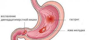
- chronic inflammatory processes in the duodenum (chronic duodenitis, peptic ulcer, Crohn's disease);
- hereditary predisposition (polyps in the intestines are the first elements prone to malignancy);
- unhealthy diet (abuse of fatty, fried, smoked foods, as well as foods containing large amounts of dyes. Deficiency of vegetables and fruits rich in fiber);
- excessive consumption of alcoholic beverages, smoking. The negative effects of alcohols and nicotine on the cells of the digestive system have been proven;
- benign neoplasms that were not treated in a timely manner;
- other diseases of the gastrointestinal tract: cholelithiasis, intestinal enteropathies, Gardner's disease, diabetes mellitus, pancreatitis and others;
- viral diseases. Some of the viruses are capable of changing the cellular genome;
- decrease in the body's defenses. When the cell's immunity decreases, lymphocytes are not able to fight cancer cells.
Classification
A cancerous tumor in the duodenum is not so rare. Most often it grows into it from the stomach or other organs. Based on the growth pattern, exophytic and endophytic tumors are distinguished. Exophytic are those that grow into the lumen of a hollow organ, endophytic beyond its boundaries.
Based on the results of histology, the disease can be divided into the following types:
- mucous cancer (adenocarcinoma). Formed from mucosal cells;
- signet ring cancer (signet ring cell). A large amount of mucin accumulates in the cells;
- adenogenic cancer. Cancer cells are very different from healthy cells;
- lymphosarcoma. A malignant neoplasm that appears from lymphatic cells;
- leiomyosarcoma. Malignant tumor of smooth muscle cells;
- neurilemmoma. A neoplasm formed by auxiliary cells of the nervous tissue;
- fibrosarcoma. Formed from connective tissue.
In case of duodenal cancer, mucinous adenocarcinoma is most often diagnosed (about 80%).
Based on location, there are 3 types of tumors:
- a tumor that is located in the descending part of the duodenum (periampullary cancer);
- cancer localized in the upper horizontal region of the intestine (suprapapillary cancer);
- a neoplasm localized in the lower horizontal region of the intestine (prejejunal cancer).
Most often, any tumor is localized where there is a greater likelihood of irritation of the organ mucosa by an aggressive environment (stomach and pancreatic juice, bile).
Like any other cancer, duodenal cancer is divided into several stages:
- Stage I. Neoplasm of small size. It grows from the mucous and submucosal layers. There are no metastases;
- Stage II. The tumor-like neoplasm begins to grow into the muscle layer, without affecting neighboring organs.
- Stage III. The neoplasm is large in size, extending beyond the duodenum.
- Stage IV. A cancerous tumor can be of any size. A distinctive feature is the formation of metastases.
Duodenal cancer: early and late stage symptoms
In older people, the development of duodenal cancer is one of the most common and dangerous cancers. The disease progresses rapidly, so it is important to make a correct diagnosis in a timely manner and begin proper treatment.
Etiology of the disease
In most cases, the tumor develops in the papillary region. In the upper intestine, neoplasms appear only in 16% of cases.
Oncology of the duodenum in exceptional cases leads to metastases, so patients have a certain chance of recovery.
If only one organ and adjacent lymph nodes are affected, there is a good chance of recovery with proper timely treatment.
Typically, recovery occurs only at the initial stage of the disease. In addition, duodenal cancer sometimes results from cancer of the stomach or pancreas.
Causes
To date, the cause of duodenal tumors has not been identified. There are certain factors that increase the risk of developing pathological processes.
- Crohn's disease. The chronic disease affects the gastrointestinal tract. In the future, there is an increased risk of developing an oncological process.
- Hereditary factor. Intestinal polyps are often inherited, which can lead to the proliferation of malignant cells. There is also an increased risk if close relatives have had cancer.
- Villous (villous) adenomas. Such neoplasms are benign, but they can lead to cancer. Adenomas have a soft consistency and can reach large sizes.
- Bad habits. Often sick people suffer from bad habits that negatively affect their overall health and lead to deterioration in well-being.
- Unbalanced and irrational nutrition. The diet often includes salty, smoked foods and avoids vegetables and fruits. Such dietary changes negatively affect the gastrointestinal tract and can sooner or later lead to cancer.
The above reasons for the development of oncological processes are the main ones, so it is important to understand the need for correct treatment measures.
Classification
When signs of duodenal cancer appear, you need to understand that oncology can develop in different ways. In each case, there is an increased risk of unwanted symptoms.
By type of growth
Tumor growth can occur in different ways:
- exophytic: the tumor grows into the intestinal lumen;
- endophytic: grows outside the intestine.
Regardless of the direction of growth, there are serious health risks.
By localization
Based on location, the tumor is divided into 3 main types:
- Periampullary cancer. In this case, the tumor is located in the descending colon.
- Suprapapillary cancer. The neoplasm is located in the upper horizontal region of the intestine.
- Prejejunal cancer. The tumor is located at the bottom of the intestine.
In most cases, the tumor is located where there is increased irritability of the mucous membrane with bile, as well as gastric and pancreatic juice.
General clinical manifestations
With the development of duodenal cancer, the first symptoms initially practically do not appear. In the early stages of oncology, it is difficult to diagnose the disease, as a result of which the diagnosis is difficult to make and treatment often begins late.
- A tumor in the papillary region of the intestine often occurs without pronounced symptoms. The diagnosis can only be made during a routine examination or in the later stages when symptoms begin to appear. People suffer from obstructed flow of bile into the intestines, pain in the right hypochondrium, nausea and lack of appetite. As the disease progresses, the skin and mucous membranes become yellow, and their shade changes gradually. Sooner or later, pancreatitis develops, since the outflow of bile is disrupted and the gastrointestinal tract functions poorly.
- Sometimes the tumor is located in the upper or lower horizontal part of the duodenum. With this type of duodenal cancer, the symptoms will be different. Intestinal stenosis may manifest itself: nausea, the stomach becomes bloated, pain appears in the right side under the ribs, heartburn, belching with sour contents, a feeling of heaviness. The development of pathology leads to intestinal obstruction, and the patient suffers from vomiting mixed with bile and a feeling of compression in the right hypochondrium.
Also, the symptoms of duodenal cancer resemble those of other cancers:
- anemia;
- lack of appetite;
- rapid weight loss without any obvious reasons for this;
- apathetic state and feeling of constant fatigue;
- decreased performance;
- increased body temperature;
- white coating on the tongue;
- profuse sweating.
Lack of appetite is a symptom of duodenal cancer
In later stages, the abdomen becomes large due to the tumor. With oncology, tumor decay may also begin, which will lead to blackening of the stool during bowel movements.
Stages of the disease
Duodenal cancer develops in stages, with each stage having certain symptoms.
- At the first stage, a small tumor is observed, and it is separated from other tissues, located in the thickness of the mucous membrane and submucosal layer of the duodenum. Symptoms may be practically absent, making timely diagnosis and further initiation of treatment difficult.
- At the second stage, a tumor develops that grows into the muscle layers of the intestine. However, there is no cohesion with neighboring organs. Single metastases appear in the nearest regional lymph nodes, which reduce the chances of recovery.
- At the third stage, the size of the tumor becomes significant. The neoplasm extends beyond the wall of the duodenum and it can grow into neighboring organs. Sometimes the tumor size remains small, but numerous regional metastases appear.
- At the fourth stage, the size and nature of the tumor can be any. Sometimes distant metastases appear.
The severity of cancer is assessed by the size and extent of the tumor, the presence or absence of metastases. For example, with cancer of the duodenal bulbs the prognosis may be more unfavorable than with other processes.
Diagnostics
Diagnosis is mandatory before starting treatment. However, people usually think about getting tested only when symptoms of cancer appear, and it may not be the first stage.
- Initially, the doctor will refer the patient for an x-ray. A bowel scan will help determine the exact location of the tumor.
- An ultrasound examination of the organ is required to determine changes in the intestinal mucous epithelium.
- They also take blood, stool, and urine tests. These examinations allow you to correctly determine the characteristics of your health condition and begin the correct comprehensive treatment.
- In many situations, it is recommended to undergo a CT scan or MRI to properly assess your health.
- A biopsy is recommended. In this case, a small piece of the formation is examined to determine the structural features.
- Endoscopy is a procedure in which a biochemical analysis is taken. This diagnostic method allows you to determine the condition of the stomach, duodenum, bile ducts, and esophagus.
- They also donate markers for oncology, since in case of tumors of the duodenum and other intestinal diseases, tumor markers for cancer make it possible to find out an accurate diagnosis.
EGDS is a method for diagnosing duodenal cancer
A comprehensive examination is required to begin successful treatment of the disease.
Treatment options
Treatment must be comprehensive. At an early stage of the disease, there is a good chance of recovery.
Surgical intervention
Surgery is required to remove the tumor. The duodenum is partially removed.
The operation is performed in patients under 75 years of age in the absence of metastases or severe chronic diseases.
Surgical intervention can be carried out according to different schemes.
- The most difficult operation is performed in cases where the tumor spreads to the duodenal papilla and the common excretory duct. In this case, part of the duodenum, head and pancreatic duct is removed. Otherwise, the operation will be ineffective.
- Circular resection is performed for small tumors. In this case, the affected area is removed. In the future, the function of the organ can be restored.
The method of surgical intervention is determined individually, taking into account the patient’s health condition.
Chemotherapy
This treatment is carried out before or after surgery. Special drugs are used to destroy tumor cells.
Radiation therapy
The method usually helps in the early stages of the disease. Radiation therapy blunts the growth of atypical cells and helps maintain the patient's condition at a certain level.
Radiation therapy helps treat duodenal cancer in early stages
Drug treatment
Drugs are prescribed for two purposes:
- reducing the risk of developing metastases and the appearance of new malignant cells;
- increasing immunity so that the body fights the disease.
Drugs are selected individually, taking into account the patient’s health condition. When carrying out treatment, you cannot use folk remedies, as they will be ineffective or turn out to be dangerous.
Palliative treatment
Palliative care allows you to improve your condition and prolong life, despite the incurability of the disease. For this purpose, symptomatic and supportive therapy is used. In difficult situations, patients are forced to turn to a hospice, where round-the-clock monitoring will be organized.
In the later stages of cancer, it is important to properly carry out pain relief, supportive and therapeutic, and immunostimulating therapy.
Forecast
It is important to know how long people live with duodenal cancer and what life expectancy depends on, how long a person can live after diagnosis.
If cancer was detected at stages 1 and 2, survival rate in the next 5 years is 70%. In other cases, survival rate is 15-20%.
The forecast depends on the following factors:
- type of neoplasm;
- presence or absence of metastases;
- presence of chronic diseases;
- patient's age.
It is advisable to make a diagnosis as early as possible to ensure effective treatment. Late stages of the disease are difficult to treat.
Prevention measures
There is no specific prevention. It is recommended to adhere to basic preventive measures:
- Be sure to give up alcohol and smoking.
- It is recommended to eat a rational and balanced diet, since fatty, high-calorie, smoked foods lead to overload of the intestines and stomach.
- Gastrointestinal diseases that can lead to cancer are recommended to be treated in a timely manner.
- Preventive examinations by a gastroenterologist are considered important for people over 50 years of age and with gastrointestinal diseases. Surveys are carried out once a year.
Preventive measures are mandatory for those who are concerned about their health and want to avoid duodenal cancer.
Source: https://oonkologii.ru/rak-dvenadtsatiperstnoj-kishki-01/
Symptoms of the disease
The clinical picture of duodenal cancer depends on the stage of the disease. In the initial stages, the disease may not manifest itself at all, or some symptoms may be confused with other gastrointestinal pathologies. After some time, aching pain in the epigastric region and a feeling of heaviness in the lower abdomen begin to appear. A characteristic symptom of duodenal cancer is “hunger pain,” which occurs when a person feels hungry.
In the later stages of the disease, the clinical picture is more pronounced. Severe intoxication of the body develops, manifested in the form of:
- chronic weakness and drowsiness. There are frequent cases of depression, apathy, life rhythm disorders, and headaches;
- a burning sensation behind the sternum and the release of gases from the esophagus and stomach;
- pallor and cyanosis of the skin. Jaundice may occur;
- dry mucous membranes and whitish coating on the tongue;
- periodic increases in body temperature;
- sudden pain in the right hypochondrium. This symptom is often confused with liver disease;
- increased sweating, especially during sleep;
- daily vomiting that does not bring relief and a persistent feeling of nausea;
- when the tumor ulcerates, bleeding begins, in which o and milena (black loose stools);
- anemia (anemia).
During the period of tumor growth in the lumen of the duodenum, changes in the pancreas in the form of inflammation are also observed. Often patients can be diagnosed with pancreatitis of varying severity or pancreatic necrosis with the formation of peritonitis.
Duodenal cancer. survival rate – Cancer Info
Duodenal cancer is a malignant tumor located in the initial part of the small intestine. This pathology is rarely diagnosed at a young age.
It most often affects men and women over 50 years of age.
Duodenal cancer: location, symptoms and signs
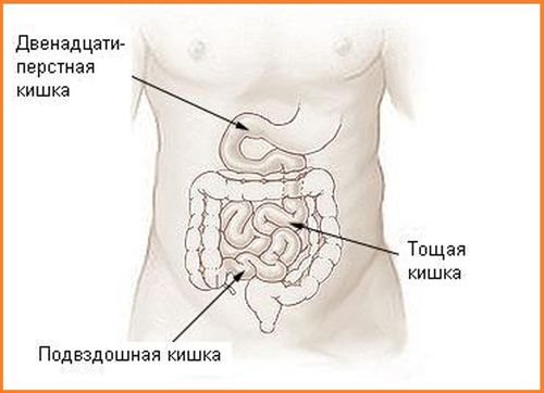
Structure of the 12 duodenum
Small intestine cancer is the development of an oncological tumor in parts of the small intestine:
- duodenum (12 duodenum);
- ileal;
- jejunum.
Cancer of the duodenum is equally common in men and women, and cancer of the jejunum and ileum is rare.
The epithelium of the duodenal glands and intestinal crypts is the site of dislocation of the cancer cell, less often the surface epithelium. Therefore, duodenal cancer is similar (duodenal cancer) to the manifestations of malignant tumors in the major duodenal papilla, head of the pancreas and common bile duct.
Cancer of the intestines and stomach metastasizes more often than duodenal cancer; the first symptoms of its metastases appear in 15-20% of patients in the regional lymph nodes near the head of the pancreas and the porta hepatis.
With the development of oncology of the duodenum, it is rarely possible to notice the symptoms of an intestinal tumor at an early stage, as well as the generalization of cancer throughout the peritoneum with hematogenous metastases.
However, there have been cases of germination of multiple metastases with a small primary tumor of the duodenum in the head of the pancreas against the background of obstructive jaundice.
It is almost impossible to diagnose duodenal cancer in the early stages; symptoms of an oncological tumor do not appear on its mucous membrane and the patient does not complain of anything. A tumor can develop in various places. As the tumor enlarges, general signs of duodenal cancer appear:
- gradual development of anemia;
- loss of appetite and weight loss;
- decreased ability to work.
As the cancer grows, it engulfs almost the entire intestinal wall and narrows the intestinal lumen. Peripapillary tumors located next to the large duodenal nipple also narrow the lumen of the bile duct and nipple.
They appear earlier than neoplasms of the duodenum in other areas, as the wall and nipple become involved in infiltrating growth. In this case, the duct above the tumor expands, bile is delayed or poorly flows into the intestines.
In this case, duodenal cancer is manifested by the following symptoms and signs:
- pain in the epigastric area and under the ribs on the right;
- nausea, decreased and loss of appetite;
- development of jaundice.
If there are no diagnostic indications indicating liver and gallbladder disease, then these symptoms indicate intestinal cancer. They are attributed to the manifestation of an oncological tumor in the large duodenal nipple.
During this period, it is extremely difficult to carry out differential diagnostics and find out where oncological diseases originated.
Tumors can be in the duodenum itself, the duodenal nipple, the common bile duct or in the head of the pancreas
During the growth of a tumor of the duodenum, the pancreas also changes and becomes inflamed, which is manifested by pancreatitis of varying severity or pancreatic necrosis with the presence of peritonitis. The symptomatology of stenosis can manifest itself in oncological tumors in the suprapapillary and upper horizontal parts of the duodenum. The scirrhous form of the tumor gradually deforms and narrows the lumen in the upper horizontal zone of the intestine.
In this case, patients will complain of symptoms similar to stenosis of the pyloroduodenal zone against the background of peptic ulcer:
- stomach discomfort;
- nausea and vomiting;
- dull persistent pain under the right ribs or in the solar plexus area;
- bloating and intestinal dilatation;
- increased acidity, which is manifested by belching or heartburn.
If the patient has already been treated for a gastric or duodenal ulcer, then stenosis is considered a complication of the ulcer. If diseases of the stomach or duodenum, including ulcers, have not been observed during life, then duodenal cancer is suspected.
It is not always possible to palpate a tumor, especially a creeping one.
With the above symptoms, colon cancer is often suspected, in particular transverse colon cancer due to its close location to the duodenum.
Causes of cancer and precancerous diseases of the duodenum
The causes of colon cancer are still not fully understood. Researchers can only speculate about risk factors for cancer, including colorectal cancer. This diagnosis combines colon cancer: rectal cancer, sigmoid colon cancer and cecal cancer.
The intestine begins with the duodenum and risk factors for its oncology include:
- chronic inflammation of the gastrointestinal tract - Crohn's disease;
- diffuse intestinal polyposis, inherited. These are small tumor-like growths of cells in the form of polyps of different shapes and sizes that protrude into the intestinal lumen;
- benign soft villous adenomas in the form of cauliflower, reaching large sizes;
- heredity: in the presence of cancer in relatives;
- abuse of spicy, salty and smoked foods, coffee, meat, animal fats and bad habits: alcohol and smoking;
- poor nutrition with a lack of vegetable and fruit products;
- diabetes mellitus, chronic pancreatitis, cholelithiasis.
Precancerous diseases of the duodenum
There are conflicting opinions among doctors about ulcerative disease of the duodenum, as its precancerous condition.
Patients complain of pain in the upper abdomen, radiating to the hypochondrium on the right and left, in the lumbar and thoracic spine, scapula and chest on the left.
The pain is accompanied by heartburn, nausea and vomiting, sour belching and a tendency to constipation. In terms of symptoms, duodenal cancer is similar in appearance to an ulcer, so it is difficult to distinguish between these diseases.
Causes and precancerous diseases of duodenal cancer
Doctors believe that in order for an ulcer to degenerate into cancer, appropriate conditions are necessary, such as long-term relapses of the disease in the absence of adequate therapy, and genetic predisposition.
It is believed that an ulcer gives rise to cancer, but it can also occur against its background.
A possible reason for the degeneration of an ulcer into cancer may be a disrupted process of reproduction and growth of cells (maturation) in the focus of inflammation of a chronic ulcer.
Other authors believe that the tendency for duodenal ulcers to degenerate (malignize) into cancer is minimal, as is the case for benign polyps (up to 15%). With multiple polyposis, the likelihood of developing into cancer increases to 30%.
If you suspect intestinal cancer, in particular the duodenum, it is important to identify in patients not only precancerous diseases, but also precancerous changes in the digestive organs as a whole. For example, you should worry if you have chronic diseases with metaplasia and dysplasia:
Therefore, a histological study of biopsy of the mucous membranes in the esophagus, stomach, duodenum, colon and small intestine, as well as biopsy of the pancreas and liver is carried out. A marker of possible malignancy in the digestive organs is liver cell dysplasia from a biopsy specimen.
Types and forms of duodenal cancer
Primary duodenal cancer is rare and accounts for 0.5% of all cases of cancer. Due to the proliferation of tumors from neighboring organs, secondary duodenal cancer may develop.
According to the forms (type of growth), cancer occurs:
- exophytic – growing into the intestinal lumen;
- endophytic - growing outside the duodenum.
Adenocarcinoma is more often diagnosed, and signet ring cell carcinoma, undifferentiated, is less common. Exocrine cancer, affecting the middle third of the duodenum, accounts for 65-75% of cases. In this case, the tumor affects the intestinal walls, narrowing their lumen, which leads to intestinal obstruction.
When a node appears that grows outward, intestinal bleeding is possible, especially with primary cancer. Some duodenal tumors cause obstructive jaundice, 10-30% spread metastases to nearby lymph nodes.
There are three tumor localizations in the duodenum:
- descending section and peripapillary region, which refers to periampullary and peripapillary cancer of the major papilla (in 75% of cases), arising from the epithelium of the pancreas or bile duct;
- upper horizontal part of the duodenum (suprapapillary cancer, 16%);
- lower horizontal part of the duodenum (infrapapillary or prejejunal cancer, 9%).
Other types of duodenal cancer include:
- lymphosarcoma arising from cancer cells of lymphatic tissue;
- leiomyosarcoma growing from smooth muscle;
- fibrosarcoma from connective tissue cells;
- malignant neuroma - from the nerve sheath.
Stages of duodenal cancer
According to the Clinical Classification of Tumors (described in the article Kidney cancer - classification, types, types, forms) duodenal cancer has 4 stages:
- Stage 1 duodenal cancer is small in size and clearly demarcated from other tissues. It is located inside the mucous membrane and in the submucosal layer of the duodenum. Regional metastases and new foci of cancer cells are not observed.
- Stage 2 duodenal cancer increases in size from 2 to 5 cm, grows into the layers of duodenal muscles, but does not damage neighboring organs. Gives single metastases to regional lymph nodes.
- At stage 3, the tumor acquires significant dimensions (more than 5 cm), extends beyond the intestinal wall and grows into neighboring organs. May be less than 5 cm, but produce multiple regional metastases.
- Stage 4 of duodenal cancer acquires different sizes and character. Has distant metastases.
TNM classification
The severity of the tumor process is assessed according to several criteria (size and extent of the tumor, metastases in the lymph nodes and distant organs). For this purpose, the TNM classification is used (Tumor (tumor) Nodulus (node) Metastasis (metastasis (spread) to other organs)).
T - size and extent of duodenal tumor:
- T1 - the tumor begins to grow through the inner wall of the duodenum;
- T2 - the tumor begins to grow into the muscle layer of the duodenal wall;
- T3 - the tumor begins to grow through the superficial membrane of the duodenum;
- T4 - the tumor has completely grown through the wall of the duodenum.
N - presence of cancer cells in the lymph nodes:
- N0 - there are no cancer cells in the lymph nodes;
- N1 - cancer cells are found in 1-2 lymph nodes near the duodenum;
- N3 - cancer cells are found in 3-6 adjacent lymph nodes.
- M0 - cancer has not spread to other organs;
- M1 - cancer has spread to areas distant from the 12th.
Diagnosis of duodenal cancer
How to identify bowel cancer? Initially, it is necessary to analyze the family history for the presence of cancer and diseases of the gastrointestinal tract. Then the patient’s life history is examined: diseases and bad habits and nutrition.
To confirm intestinal cancer, diagnosis of the skin, mucous membranes of all organs: mouth and nose, eyes is necessary to confirm jaundice and cachexia (exhaustion).
Also, a blood test for intestinal cancer is of great importance in diagnosis: anemia (anemia) is determined by a general blood test. As a result of laboratory tests:
- from the blood in case of intestinal oncology helps to identify tumor markers - special proteins that are secreted by tumors; a detailed analysis - increased levels of albumin (protein);
- A general urine test can also determine protein (proteinuria) and red blood cells (erythrocyturia);
- A stool test detects blood in the stool.
Colonoscopy, as an instrumental method, is performed with an endoscope. At the same time, the internal state of the intestines is examined and assessed.
How to check the intestines for cancer without a colonoscopy:
During irrigoscopy, X-rays and a contrast agent are used, and the intestines are first cleaned. The study will be carried out in two ways: the stomach and duodenum. A video camera is built into the endocapsule to study the structure of the gastrointestinal tract and detect pathology.
This type of diagnosis is carried out for abdominal pain, hidden bleeding and suspected cancer. The patient swallows the capsule on an empty stomach.
After 8 hours, the capsule comes out with the feces, and during this time all disorders in the stomach and intestines are recorded.
Using a sigmoidoscope, the intestines are examined through the patient’s anus, then material is taken for biopsy. This is prescribed for polyps and other neoplasms in the rectum.
Computed tomography is a virtual way of performing a colonoscopy without a contract substance or insertion of instruments. The degree of compression of organs by the tumor is determined. A tumor is detected by MRI, which is a more accurate method than CT.
During endoscopy, the doctor examines the inner surface of the esophagus, stomach, duodenum with an endoscope and takes a fragment of the organ for biopsy. Ultrasound indicates the presence of a tumor and metastases.
Additionally, the chest is examined with X-ray to determine metastases and secondary cancer. Examinations are carried out by a gastroenterologist and a therapist.
Treatment methods and life expectancy for duodenal cancer
Duodenal cancer is treated with gastropancreaticoduodenectomy (GPDR). In this case, nearby lymph nodes are removed. For small tumors (less than 1 cm): endocrine cell and nonepithelial; in elderly patients with complicated diseases, the tumors are excised and part of the duodenum is removed. Surgical intervention in this case will be non-radical.
Source: https://rak-info.ru/drugoe/rak-dvenadtsatiperstnoj-kishki-vyzhivaemost.html
Diagnostic methods
The tests prescribed to make a diagnosis are based on anamnesis. The doctor needs to know the patient’s complaints, he conducts an examination and studies the test results. Almost all patients are prescribed instrumental examination methods.
Instrumental methods include:
- fibrogastroduodenoscopy;
- Ultrasound examination;
- CT scan;
- magnetic resonance imaging.
FGDS is a diagnostic method that allows you to examine the upper parts of the digestive tract, including the duodenum, using an endoscope. It can also be used to take a piece of material for a biopsy. Nowadays, it is one of the most reliable diagnostic methods and allows you to make a diagnosis in the shortest possible time. The procedure is carried out on an empty stomach (the last dose is no earlier than 12 hours), it is not recommended to take medications or smoke (nicotine promotes increased secretion of hydrochloric acid).
Before the operation, you are allowed to drink weakly brewed tea or still mineral water 2-3 hours before the operation. There are a number of contraindications to the examination: mental disorders, diseases of the larynx and pharynx in the acute stage, enlarged cervical lymph nodes, acute myocardial infarction, bleeding disorders, narrowing of the lumen of the esophagus, serious diseases of the thyroid gland, exacerbation of bronchial asthma.
There are cases when an FGDS is necessary and is carried out urgently, despite the presence of contraindications. In this case, the patient will be in a hospital setting, where, if necessary, he will be immediately provided with first aid. Complications may include bleeding, perforation of the esophagus, stomach or duodenum, or infection in the digestive tract.
Ultrasound is a diagnostic method based on the interaction of ultrasonic waves and the tissues being examined. Using ultrasound, you can identify the anatomical and functional state of the duodenum. This method is most often used as a primary diagnosis, since with its help it is not always possible to obtain complete information about the identified pathology. To obtain reliable information, saline is first injected into the stomach. solution or other liquid.
A few days before the start of the study, you need to adhere to a small diet: limit yourself to the consumption of fresh vegetables and fruits, fatty meats and fish, exclude legumes, carbonated and alcoholic drinks. Approximately 8 hours before the procedure, refuse to eat, and before starting it, drink about a liter of any liquid (water, tea, juice). The procedure usually takes no more than half an hour. There are no complications observed after the procedure.
CT and MRI are similar diagnostic methods. These methods are based on the impact of nuclear magnetic resonance on organ tissue and obtaining data on tomographic images. The study is carried out strictly on an empty stomach; even liquid intake is not recommended. Before starting the procedure, it is necessary to remove all metal devices: hearing aids, implants, remove products, rings, earrings, chains, etc. It is not allowed to undergo the procedure while the cell phone is working.
Contraindications for CT and MRI are the period of gestation, children under 7 years of age, fear of confined spaces, metal fragments that cannot be removed (dental and eye implants, pacemakers). There are practically no complications observed after the procedure. Allergic reactions to the contrast agent or damage to organs that have even minor metal elements may occur.
Laboratory methods include:
- general clinical blood test (hemoglobin level, presence of anemia, increased liver enzymes due to stagnation of bile);
- feces for occult blood (determination of bleeding at an early stage of its development);
- blood for tumor markers (special analysis to identify cancer cells).
Based on the information collected, the doctor can make the correct diagnosis and prescribe effective treatment.
Diagnosis of malignant tumors of the duodenum
Laboratory tests do not provide the doctor with any specific tests.
With cancer and sarcomas of the duodenum, a gradual decrease in the number of red blood cells and hemoglobin is observed in the blood, sometimes significant. Neutrophilic leukocytosis is often observed, especially with peripapillary tumor localization with the development of cholestasis and secondary cholangitis. ESR increased to 20-50 mm per hour. When a tumor grows into the pancreas, thrombocytosis is possible.
With the development of polestasis and jaundice, hyperbilirubinemia and a positive reaction to bile pigments and urobilin in the urine are detected. The reaction to occult blood in stool is often positive.
Examination of gastric functions in most patients reveals hyposecretion and hypoacidity of gastric juice. The lowest rates are typical for lesions of the proximal parts of the duodenum. In the duodenal contents you can find traces or a more significant content of red blood cells due to the disintegration of the tumor, a large number of leukocytes. Atypical cells may be found.
Treatment and survival prognosis
Currently, the only treatment for duodenal cancer is surgery followed by radiation and chemotherapy to prevent relapses.

Surgical treatment involves removal of a malignant tumor. In this case, part of a healthy organ is excised along with it. Surgery is performed on persons under 75 years of age, except in cases of metastases. Tumors of stages III and IV are practically not operated on due to the fact that during surgical intervention there is a decrease in immunity, and this is an impetus for the rapid growth of the tumor and the proliferation of metastases in neighboring organs and tissues.
Radiation therapy (radiotherapy) is ionizing radiation directed at the site of diseased tissue, which suppresses the growth and activity of cancer cells. Radiation therapy is of great importance - it significantly reduces the risk of metastases.
Chemotherapy is also an integral part of the treatment of cancer. The essence of the procedure is to take medications that destroy cancer cells. At the same time, the negative impact on the body as a whole is minimized. Taking medications should be carried out exclusively under the supervision of an oncologist.
Survival among all patients suffering from this pathology depends on many factors. For patients in whom cancer is detected in the first 5 years, and the cancer is diagnosed at stages I and II, the survival rate is about 75%. If stages III and IV are detected, and the tumor is removed, the survival rate will be only 20%.
It is also necessary to take into account other indicators: the patient’s age, tumor type, absence of concomitant diseases, presence or absence of metastases.
Important! Duodenal cancer is highly treatable in the early stages of its development, and in order to make a diagnosis, it is necessary to regularly undergo a medical examination.
Prevention of neoplasms
Starting from a young age, it is necessary to follow some rules that reduce the risk of developing cancer of the stomach and duodenum. It won’t be difficult to do them and they are recommended for absolutely everyone:
- Diversify your diet with vegetables and fruits;
- Eat plenty of fiber;
- Give up bad habits: drinking alcohol and smoking;
- Exercise regularly and lead a healthy lifestyle;
- If diseases of the stomach and duodenum occur, carry out timely treatment;
- Reduce the risk of constant presence of excess acid in the stomach (heartburn) by taking special medications and eating restrictions;
- Get an annual examination.
Conclusions: duodenal cancer develops very slowly. Any disease associated with the digestive system should not be ignored, since many diseases are precursors to oncology. With regular examination, there is a high probability of diagnosing a tumor at an early stage of development. It will also help to contact a specialist in a timely manner if any unpleasant symptoms appear. The survival rate of patients with stage 1 and 2 cancer is quite high. Therefore, you should not neglect treatment and take care of your health.