The study is unpleasant, but the most informative of all diagnostic measures existing today. During the examination, the doctor may remove single small polyps or take a tissue sample for a biopsy. The procedure is prescribed for chronic forms of gastritis and ulcerative pathology to confirm the diagnosis and select a treatment regimen.
METHODS OF DIAGNOSIS OF THE STOMACH
There are several methods for medical diagnosis of the condition of the gastric mucosa:
- physical – performed in the doctor’s office;
- laboratory - examine the patient’s tests;
- hardware - using medical devices.
Physical methods are a routine examination performed by a doctor. The doctor listens in detail to the person’s complaints, conducts an initial examination - the oral cavity, tongue, palpates the lymph nodes and the abdominal area.
Laboratory tests are carried out to identify the causes of stomach pathology - what concomitant diseases could provoke the disease? For diagnosis, blood, feces and urine are taken.
Hardware diagnostics include ultrasound and fluoroscopy. In modern medicine, diagnostics are used - gastropanel. This is a paid alternative to gastroscopy - a laboratory blood test.
An absolute contraindication to gastroscopy of the stomach is the patient’s near-death state. Diagnosis is possible even with a heart attack and in the presence of gastric bleeding. However, there are contraindications to the procedure:
- risk of aortic rupture;
- heart ailments - they are treated first;
- hemophilia - there is a risk of tissue injury;
- high blood pressure;
- diseases of the neck area;
- anatomical deviations in the patient's body structure.
If gastroscopy is not possible, gastric diseases are determined using alternative methods.
How to properly prepare for research?
First of all, it is recommended to seek advice from a general practitioner or gastroenterologist. They will conduct a general examination, palpation of the abdomen, and also collect anamnesis. Before undergoing this diagnostic procedure, the patient should find out what tests are needed for gastroscopy of the stomach. Most often, therapists and gastroenterologists refer a person for the following clinical and laboratory tests:
- general blood analysis;
- biochemical blood test (ALAT, AST, total and conjugated bilirubin, amylase, electrolytes);
- blood glucose.
In addition to taking tests, it is very important to undergo the necessary preparation immediately before esophagogastroduadenoscopy. It means refusing to eat any food for at least 6 hours before the test.
Carrying out capsule endoscopy requires proper preliminary preparation to facilitate the procedure. Here is a list of recommendations:
- Two days before the test, exclude alcohol and carbonated drinks, baked goods, legumes, and white cabbage from the diet.
- Stop taking iron supplements one week before.
- The day before, cleanse the intestines using the drug “Fortrans”.
- Do not eat 12 hours before the procedure.
Electrodes are placed on the patient's stomach and signals from an empty stomach are studied for 40-50 minutes. The method is aimed at studying the motor-evacuation function of the digestive tract. It is safe and easy to implement. There are no contraindications to electrogastroenterography.
During X-ray examination, contrast is performed using barium sulfate. Without it, the information content will be zero. The image allows you to assess the structure of the organ. But often this data is not enough and gastroscopy is also prescribed. One of the advantages of x-rays is that the method is painless.
It is difficult to answer this question unequivocally. If we compare these two procedures, then MRI does not show the condition of the mucous membrane, unlike gastroscopy. Also, there is no way to take a biopsy. Another disadvantage is the high cost. On the other hand, magnetic resonance imaging is a painless procedure in which the patient does not experience any discomfort. In some cases it is advisable to use it.
Ultrasound examination allows one to distinguish between neoplasms and some pathologies that cannot be detected with an endoscope. Gastroscopy helps to identify the localization of ulcerative lesions and assess the depth of damage to the mucous membrane. Ultrasound is a comfortable procedure, unlike probing.
The decision to perform gastroscopy of the stomach without swallowing the probe is always made by the attending physician. It assesses the patient’s health status, presumptive diagnosis and then selects the optimal diagnostic method.
This article is posted for educational purposes only, does not replace an appointment with a doctor and cannot be used for self-diagnosis.
Easy diagnostics
Simple diagnostic methods are mandatory for use when a patient complains of an acute abdomen, nausea and other symptoms of gastric diseases.
Physical examination
Physical measures are carried out at a doctor’s appointment, the results depend on the qualifications of the medical specialist. The complex includes:
- studying the anamnesis, assessing symptoms from the patient’s words;
- visual examination of mucous membranes;
- feeling painful areas of the body (palpation);
- palpation in a specific body position (percussion).
Based on the results obtained from such an examination, it is extremely difficult to diagnose the disease. The doctor may suspect the presence of pathology, but more in-depth research methods are needed to confirm it.
Microscopic laboratory diagnostics
Laboratory methods involve taking samples from the patient for further study and evaluation of the results. Most often, the following physical and chemical studies are prescribed:
- general urine analysis;
- coprogram (stool analysis);
- clinical blood test. The number of all types of blood cells (erythrocytes, leukocytes, platelets) is counted and the hemoglobin level is determined;
- gastropanel. This blood test is aimed at studying the condition of the gastric mucosa. Based on its results, the following is determined: the presence of antibodies to Helicobacter pylori bacteria, the level of pepsinogen proteins produced, the level of the polypeptide hormone - gastrin, with the help of which the acidic environment in the stomach is regulated;
- blood biochemistry. Quantitative indicators of bilirubin, liver enzymes, cholesterol and other blood elements are established.
Tests help identify inflammatory processes and other disorders of organs and systems. If the results differ significantly from the normative indicators, the patient is prescribed an instrumental or hardware examination.
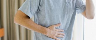
Symptoms
Stomach diseases are expressed by the following symptoms:
- Nausea is typical both before and after meals. This symptom is also often observed in the morning.
- Vomiting occurs with poisoning or a progressive ulcer. In the latter case, vomiting may be bloody.
- Sudden weight loss and pallor are a sign of the development of stomach cancer.
- Heartburn occurs after eating acidic foods.
- Decreased appetite.
- Sleep disturbance.
- The appearance of pain in the stomach. In this case, the nature of the pain can be different (aching, dull, cutting, stabbing).
- The appearance of unpleasant belching.
- Weakness.
- Indigestion.
- Abnormal stool.
- Increased body temperature.
- Taste disturbance.
- Yellowing of the skin.
- Constipation.
All of the above symptoms can develop within a few days (in acute forms of the disease).
If the pathology has already become chronic, then the patient will only be bothered by occasional nausea and abdominal pain. Typically, advanced diseases have a monotonous course with periods of exacerbations.
Laboratory methods for examining the stomach
How can you tell anything about the stomach based on blood results? It turns out that it is possible! Laboratory methods are of great importance in establishing a diagnosis and are actively used in gastroenterology. The material for the study is the patient’s blood, feces, gastric and duodenal juice.
Blood
A general and biochemical blood test is the first thing that any doctor prescribes to diagnose gastritis. Blood counts and their levels can tell a lot about your general condition, the presence of an inflammatory process, infection, and the functioning of digestive enzymes and hormones. Specifically, indicators of gastric function include hemoglobin, leukocytes, erythrocyte sedimentation rate (ESR), pepsinogen and gastrin levels. Their fluctuation indirectly indicates the presence of gastritis, ulcer bleeding and other diseases.
The amount of antibodies to Helicobacter pylori (immunoglobulins M and G) is also examined in the blood. Helicobacter is a bacterium that causes inflammation of the gastric mucosa. If it is detected, antibacterial therapy is prescribed.
Feces
Stool analysis shows the presence of hidden bleeding, enzyme dysfunction (changes in coprogram), worm eggs, and signs of dysbacteriosis.
Gastric juice
This method will tell you how to determine the functional state of the gastric mucosa. The juice is collected through a thin probe. Each portion of the material is examined separately. pH-metry is also carried out - this is a determination of acidity. The obtained indicators play an important role in prescribing treatment.
All laboratory methods are used to diagnose functional indicators of the stomach, and therefore are not suitable for determining space-occupying formations, narrowing or obstruction of the esophagus, or detecting the source of bleeding.
Which doctor examines the stomach
Intestinal problems have become increasingly common lately. Many people try to cope with them on their own, but often a situation arises when they cannot do without the help of a specialist.
Having suffered from pain and flatulence, a person wonders which doctor to see. The condition of the intestines is very important for the health of the entire body, because it not only absorbs nutrients, but also produces hormones, enzymes, vitamins, and develops immunity.
Therefore, if problems arise, you need to start treatment as soon as possible.
Intestinal structure and diseases
The intestines are the largest part of the digestive system. Its length is at least 6-7 meters.
It is in the intestines that the main process of breaking down food into nutrients, as well as their absorption, occurs.
In addition, many enzymes, hormones and vitamins necessary for the normal functioning of the body are formed here. The immunity that protects a person from infections is also formed in the intestines.
This organ consists of two sections: the small and large intestines. The small intestine starts from the stomach. It has a small diameter and thin walls.
This section begins with the duodenum, where the main processes of digestion take place. This is where bile and pancreatic enzymes come.
Most medications and many nutrients from food are also absorbed in the duodenum.
The small intestine in the pelvic area passes into the large intestine. The large intestine can have a diameter of up to 8 cm and thick walls.
This is where beneficial microorganisms live, which are responsible for human immunity and the production of many vitamins. Water, vitamins, glucose, and amino acids are absorbed in the large intestine.
This is where feces are formed. They can be retained for about a day, then are excreted through the rectum.
Due to the huge area of the intestinal walls and the functions it performs, any digestive disorders affect its condition.
Depending on the cause and nature, different intestinal diseases occur:
- Dysbacteriosis is now considered the most common pathology of the gastrointestinal tract. The normal intestinal microflora is disrupted due to uncontrolled use of NSAIDs or antibiotics, poor nutrition, stress, and frequent viral diseases. It can also occur due to chronic gastritis or allergic diseases. The passion of many people for cleansing procedures and enemas also greatly disrupts the intestinal microflora.
- Enteritis is an inflammatory bowel disease. The acute form of this pathology can occur due to poisoning, infection, overeating, or eating spicy food. Poor nutrition and stomach pathologies can lead to chronic enteritis.
- Inflammatory diseases also include intestinal colitis. This pathology is localized in the large intestine. Colitis can be ulcerative, infectious, toxic, ischemic or spastic. But regardless of the cause, the disease greatly disrupts the microflora, leads to inflammation of the mucous membrane, and impaired absorption of nutrients.
- Crohn's disease affects the entire digestive tract. This is a chronic pathology that is accompanied by nonspecific symptoms, but disrupts the functioning of the entire body. The prognosis depends on the timeliness of its diagnosis.
- Irritable bowel syndrome is said to exist if there are no inflammatory processes or neoplasms, but the functions of the organ are impaired.
- Duodenal ulcer most often occurs along with damage to the walls of the stomach. This is a chronic pathology, the cause of which may be poor nutrition, stress or hereditary predisposition. But recently it is believed that the appearance of ulcers is caused by the bacterium Helicobacter pylori.
- The most common tumors in the intestine are polyps. The cause of this pathology can be a hereditary predisposition, a sedentary lifestyle, and poor nutrition. Polyps seriously interfere with the functioning of the intestines and can lead to intestinal obstruction.
- Colon cancer most often affects the colon. Usually the disease is diagnosed only in late stages, as it does not have specific symptoms.
When to see a doctor
Even the slightest discomfort in the gastrointestinal tract cannot be ignored. Usually people who have been suffering from stomach problems for a long time try to treat themselves. But this can be dangerous, because indigestion affects the condition of the entire body.
Often, intestinal problems are a consequence of gastritis, liver or gallbladder pathologies. In this case, the patient is usually already being treated by a therapist or gastroentrologist. But it is very important to know when you need to see a doctor urgently.
Typically, intestinal problems are indicated by the following signs:
- pain during defecation;
- prolonged constipation;
- frequent diarrhea;
- blood, mucus, or undigested pieces of food in the stool;
- pain in the navel or lower abdomen;
- decreased appetite;
- nausea, vomiting;
- flatulence, bloating, increased gas formation;
- belching, heartburn;
- weight loss, metabolic disorders;
- decreased immunity;
- general weakness, decreased performance.
Which specialist should I contact?
Intestinal problems vary in origin and location. And during treatment it is very important to take this into account. Therefore, it is impossible to say exactly which doctor treats the intestines. When mild pain or diarrhea occurs, you should consult a physician.
He will prescribe an examination that will help determine the cause of the pathology and choose a treatment method. The therapist gives a referral to more specialized specialists if the pathology turns out to be serious.
It is also recommended to consult other doctors if problems with the intestines are chronic.
Source: https://limto.ru/kakoj-vrach-obsleduet-zheludok/
Ultrasound examination (ultrasound)
It is used every day as a diagnostic method for determining many diseases. There is not a single organ that cannot be examined using ultrasound, but more often they examine dense structures and tissues. As for hollow organs, such as the stomach, not everything can be seen there. For example, it will not be possible to determine the types of gastritis, but it is possible to detect a neoplasm, polyp, or change in shape. Thus, if a doctor is faced with a choice of what is better to do: ultrasound or fibrogastroscopy, the answer is obvious! FGS will give the best result.
Magnetic resonance imaging (MRI) and computed tomography (CT)
To examine the stomach and esophagus, MRI and CT are used very rarely, but they are quite applicable as an alternative method. The principle of operation of modern computed tomographs is based on emitting a magnetic field on the body and receiving impulses from internal organs, which are recorded on a special film in the form of clear images. The step of obtaining sections (images) is set by the program or the doctor. Thanks to the MRI machine, you can thoroughly study every millimeter of any organ. CT also examines organs layer by layer, only using x-rays.
Today, this examination is considered the most accurate and safe, since doctors have a real opportunity to look inside the body without intervention and examine any structure. As you may have guessed, tomography also has contraindications and disadvantages. A relative contraindication is pregnancy. The disadvantages of MRI include the inability to examine the function of an organ, monitor its functioning, secretory and enzymatic activity. CT is used even less frequently.
You cannot examine a patient with any metal structures in the body (bone pins, screws, vascular clips), and especially with pacemakers. Failure to comply with this condition may cost a person his life.
Not long ago, 2 completely new methods of diagnosing the stomach without the use of a fiberscope and the associated discomfort appeared: gastropanel and capsule gastroscopy.
What is better: gastroscopy or x-ray?
Contrast X-ray of the stomach is the most common diagnostic method in gastroenterology.
The accuracy of this technique is about 75%. With the classic procedure, you can check the following:
- state of gastric motor function;
- relief of the mucous membrane;
- shape and contours of the stomach.
If necessary, double contrast x-rays are used. In this case, contrast (barium sulfate) and air are used. This method allows you to diagnose diseases in 90% of cases.
X-ray examination with contrast can detect various diseases of the stomach - for example, pyloric stenosis
X-ray diagnostics are contraindicated in pregnant women, especially in the first trimester. Contrast is not used if perforation of the gastric wall is suspected and after surgery.
Using this technique, the following diseases can be identified:
- gastritis without specifying the clinical form;
- stomach polyp;
- ulcer and its complications;
- stomach cancer.
The final diagnosis is not always made by x-ray examination. Often only indirect signs of the disease are detected.
With radiography, it is not possible to visually assess the condition of the mucous membrane or see signs of inflammation. Therefore, such diagnostics are not a complete replacement for fibrogastroscopy.
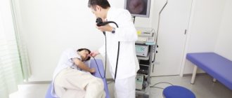
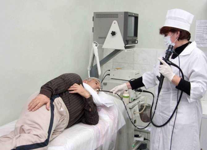
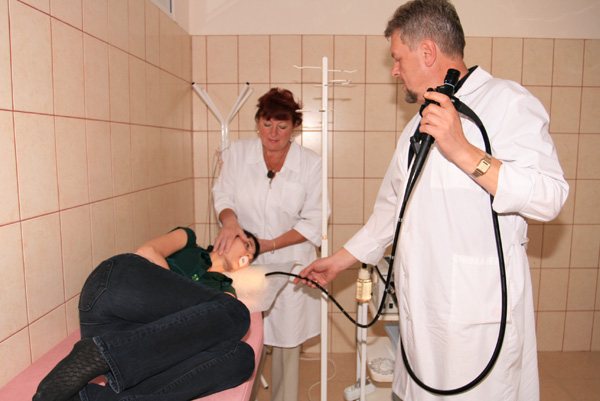
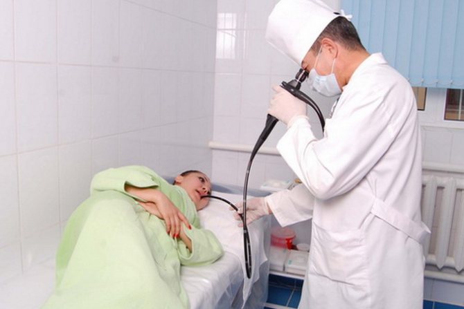
Let us remind you that gastroscopy of the stomach is a type of endoscopic examination in which, using a special device - an endoscope - the doctor examines the inner surface of the stomach.
That is, the doctor sees the entire internal surface of your organs with your own eyes in an enlarged form on the monitor screen. This is similar to going on a tour and seeing the sights with your own eyes.
All other diagnostic methods in this case are something akin to a photocopy of a photograph that was not even taken by you yourself.
| The gastric mucosa is normal. Visual picture during gastroscopy | And this is what we see on an X-ray of the stomach with barium contrast |
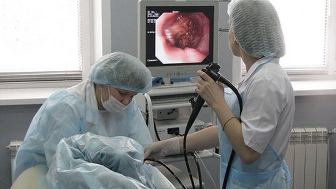
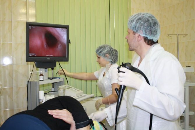
With gastroscopy, the doctor sees the condition of your organ from the inside in real time, with magnification and good lighting. This allows you to consider what changes there are in the stomach, whether there are neoplasms (even at the very initial stage), erosions, ulcers, polyps, etc.
If there is any formation, then you can immediately feel it (palpation with an instrument) and immediately take a biopsy.
This allows you to make an accurate diagnosis and immediately prescribe the necessary treatment.
If you first do an x-ray of the stomach, then if it shows any deviations from the norm, then you will still be sent for a gastroscopy to understand in detail what’s what.
For example, an x-ray showed “filling defects” - this is when the contrast agent (barium) does not fill any areas in the organs. There is an assumption about the presence of an ulcer or neoplasm. And to really understand what is there, after the x-ray you will be sent for a gastroscopy.
An X-ray of the stomach (necessarily with barium) is more of an additional examination than the main one. Most often it is prescribed:
- to detect narrowing of the esophagus, stomach, duodenum;
- to identify protrusions (diverticula);
- with reflux into the esophagus (reflux);
- in the presence of bile in the stomach (gastro-duodenal reflux - from the duodenum to the stomach);
- to detect deformations of the duodenum;
- track the dynamics of food movement through the gastrointestinal tract (GIT) over time and evaluate the motility of the GIT, detect intestinal obstruction.
Also, x-rays can be an aid in diagnosis when, due to absolute contraindications, gastroscopy cannot be done.
MRI (magnetic resonance imaging) is an examination in which we obtain a layer-by-layer (tomography) image of the internal structure of the human body. As a rule, one section is targeted - the chest, abdominal cavity, head, etc.
The name of the method itself is based on the principle of obtaining such sections - magnetic resonance of living tissues.
CT (computed tomography) is also an examination in which we can obtain layer-by-layer sections of the internal structure of a person, just like MRI.
The main difference is the principle of obtaining slices (X-rays) and the assistance of a computer in creating images.
Both types of examination appeared relatively recently and, like everything modern, they are immediately classified as better and more progressive. But it is not always the case.
Often, good old, proven methods are much more informative and reliable than new ones. If only because they have accumulated a lot of experience in the variety of clinical pictures and their interpretation, there are schools in which the experience of interpretation and diagnosis is passed on by word of mouth - from an experienced doctor to a young specialist.
To read MRI and CT images correctly, a lot of experience and time is required, which the doctor can devote to studying the results of the examination of one patient (at least 1 hour to look at all the images and make a conclusion). On the fly, the doctor, as a rule, does not have such time.
A CT scan is one of many cross-sections taken at the level of the abdominal cavity.
The arrow shows the stomach
Advantages of the CT method:
- allows you to study the functioning of organs using intravenous administration of a contrast agent - measure the speed of blood flow, cerebrospinal fluid flow, etc.
- Thanks to a new generation of high-resolution CT scans and innovative software, it has become possible to construct 3D models of all organs and systems of the body.
CT comes to the rescue in cases where endoscopic examination is not possible. For example, with severe pathologies of the cardiovascular and respiratory systems.
Gastropanel
This blood test can check several indicators. Based on their level, an experienced and competent doctor will be able to draw conclusions regarding the pathology of the gastric mucosa.
So, the gastropanel explores:
- Antibodies to H. pylori (Helicobacter), the bacteria that causes stomach ulcers.
Antibodies to Helicobacter pylori of the IgG class are detected starting 3 weeks after infection
- Pepsinogen I and II (precursors of the gastric enzyme pepsin). By their meaning one can judge which part of the stomach is affected.
- Gastrin 17 (a hormone that regulates the production of hydrochloric acid).
Based on the totality of all indicators, a conclusion is drawn in which the doctor indicates the degree of dysfunction of the gastric mucosa and possible causes (atrophy, hypotrophy, hyperacidity and others). The examination is expensive and not informative enough, since it is impossible to visually see the condition of the organ from the inside, but sometimes it is an excellent diagnostic method stomach without gastroscopy.
Many diseases, including cancer of the stomach or esophagus, may not manifest themselves and are detected at the last stage.
That is why gastroscopy remains the leading examination for diagnosis.
Capsule gastroscopy
A new type of examination of the esophagus and stomach from the inside using photographic equipment. We can say that this is a good alternative to gastroscopy of the stomach. The device is a small capsule (10 mm) with a built-in lens that allows you to take many pictures as it progresses. Like any other study, it is carried out on an empty stomach. The patient washes the capsule down with water and can do his usual activities. After 8-9 hours the capsule comes out naturally. Reviewing all the information recorded during this time, the doctor draws conclusions about the condition of the examined gastrointestinal mucosa and the detected formations.
It is worth saying that capsule gastroscopy is not used in all medical institutions due to its high cost. This survey is currently at the innovation stage. Therefore, for a high-quality diagnosis of gastrointestinal diseases, every doctor needs to think not what can replace gastroscopy, but how to overcome his fear and set him up for the necessary examination.
SOURCES: https://diagnostinfo.ru/skopiya/gastroscopy/kak-proverit-zheludok-bez-gastroskopii.html https://apkhleb.ru/endoskopiya/obsledovanie-zheludka-bez-gastroskopii https://proskopiyu.ru/ gastroskopiya/kak-mozhno-proverit-zheludok-bez-gastroskopii.html