Diagnosis of intestinal diseases is based not only on an analysis of the patient’s complaints and characteristic symptoms, but also on data from special examinations. Instrumental studies are crucial in making an accurate diagnosis. Depending on the characteristics of the clinical picture of the disease, various examination methods can be used in patients.
History taking
Diagnosis of intestinal diseases begins with collecting anamnesis - questioning the patient. The patient needs to describe in detail all the symptoms that are presumably caused by pathology of the digestive system. Intestinal damage can be suspected if the following signs appear:
- diarrhea;
- constipation;
- flatulence and bloating;
- pain in the lower abdomen.
The doctor asks the patient in detail about the nature of the symptoms. The time of occurrence of disturbances (connection with food intake, time of day) is of great importance. When collecting anamnesis, the dynamics of the development of symptoms is monitored in detail - whether they intensified over time, or whether new signs of pathology appeared.
A complete clinical picture of the disease allows the doctor to suspect the presence of the disease in the patient. Based on the medical history, a preliminary diagnosis is made, which must be confirmed using instrumental diagnostic techniques.
Without additional research, it is impossible to accurately determine the disease and prescribe treatment to the patient. Only a full range of diagnostic procedures will allow us to establish the cause of symptoms, according to which treatment will be selected.
Laboratory examination methods
If the doctor suspects the development of intestinal pathologies, the examination begins with a blood, urine and stool test. The results of such studies will confirm or refute the presence of pathological processes in the body.
A general blood test is prescribed for suspected inflammatory and infectious processes, bleeding, parasitic lesions, and oncology. The collection of biological material is carried out exclusively with sterile instruments. In adults, blood is taken from the ring finger, and in small children, from the big toe.
Intestinal diseases affect changes in blood parameters, namely:
- red blood cells and hemoglobin. Anemia may indicate the development of internal bleeding;
- lymphocytes. These cells reflect the level of activity of the immune system. During infectious and oncological processes, the level of lymphocytes can either increase (lymphocytosis) or decrease (lymphocytopenia);
- monocytes. An increase in the number of these cells indicates the development of an infectious lesion;
- eosinophils. An increase in this indicator (eosinophilia) most often indicates helminthic infestations. But this may also indicate the presence of malignant neoplasms. A decrease in the number of eosinophils is usually observed in the postoperative period, as well as in the initial stages of the infectious process;
- erythrocyte sedimentation rate (ESR). An increase in this indicator may indicate inflammatory and infectious processes.
Physical examination techniques
Physical techniques are studies that a doctor can conduct without the use of special equipment. These include a general examination of the abdomen, palpation and rectal examination. In rare cases, percussion and auscultation are used.
Examination of the patient
An examination is a visual assessment of the condition of the patient’s anterior abdominal wall. Using this technique you can evaluate:
- skin color, presence of pallor, cyanosis or local redness;
- elasticity, skin turgor;
- increased muscle tone of the abdominal wall is one of the symptoms of peritonitis;
- the presence of skin rashes, roughness of the integument.
An indirect symptom of disorders in the intestines can also be a varnished surface of the tongue or the appearance of a white coating on its surface.
We recommend reading:
Preparation and conduction of FGDS (fibrogastroduodenoscopy)
Palpation of the abdomen
Palpation is palpation of the abdomen, assessing the condition of the anterior abdominal wall. Depending on the depth of pressure, superficial and deep palpation are detected.
First, the patient undergoes superficial palpation of the abdomen. The doctor consistently presses on the surface of the abdominal wall, moving in a circle. The patient must relax the abdominal muscles. Signs of dysfunction of the digestive system, which can be detected by palpation, are:
- increased or weakened intestinal motility (strength of muscle contractions of the organ);
- the presence of pain limited to a certain area or spread over the entire surface of the abdomen;
- increased tone of the abdominal muscles - rigidity, indicating the spread of the process to the peritoneum.
In the place where pathological changes are determined, the doctor performs deep palpation. It involves pressing the fingers firmly into the abdominal wall, while the patient must exhale. When using deep palpation, the doctor can more accurately assess changes characteristic of diseases of the digestive system.
Rectal examination
A rectal examination is palpation of the anus and anal canal in order to assess their functional activity. The examination is carried out with the patient lying on his side with his legs drawn to his body and his knees bent.
A rectal examination is performed if the patient has local symptoms (itching and burning in the anal area, bleeding). Diagnosis is the first step in identifying such a common disease as hemorrhoids.
Read on the topic: Using a rapid test from a pharmacy to determine occult blood in stool
Are there alternative methods to check the intestines without colonoscopy?
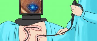
Those who have already encountered this unpleasant procedure are wondering how to check the intestines without a colonoscopy? After all, this diagnostic method not only gives the patient a lot of unpleasant sensations, but also requires lengthy and complex preparation. The effectiveness of the procedure is invaluable, but still patients prefer to look for an alternative. Modern medicine offers them other diagnostic methods, which in some cases can actually replace colonoscopy.
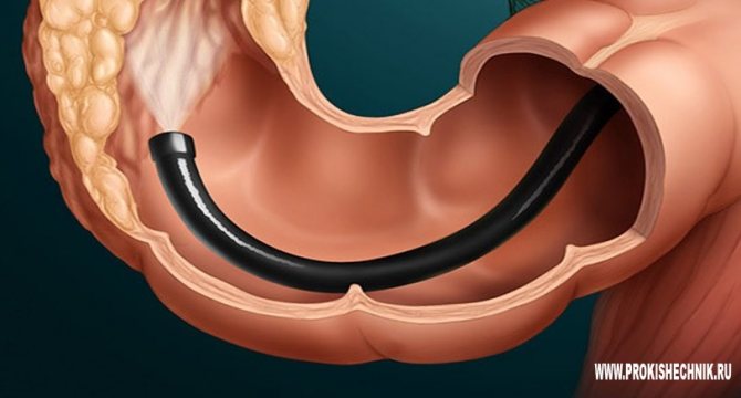
What is a colonoscopy?
During the examination, a special flexible tube with a camera located at its end is inserted through the rectum.
Thus, it is possible not only to examine the large intestine, but also to remove fecal stones or existing polyps from its walls.
Colonoscopy is not used to examine patients with cardiac, pulmonary or renal failure, with exacerbation of intestinal infection, bleeding disorders, colitis, or peritonitis.
In addition to the fact that the procedure itself causes the patient a lot of discomfort and unpleasant sensations, you still need to carefully prepare for it.
During the day before the colonoscopy, the patient must follow a liquid diet, take laxatives and do enemas. All this is necessary to completely cleanse the intestines.
How to check the intestines and how to replace this or that procedure should be decided by the doctor. When choosing a colonoscopy, the doctor will tell the patient about the advantages and disadvantages of such a diagnosis.
The positive points include:
- Today this method is considered the most effective and reliable.
- Thanks to wide visualization, the condition of the organ can be fully assessed.
- During the procedure, it is possible to remove polyps and stop bleeding without resorting to surgery.
- Diagnostics takes no more than 30 minutes.
- The doctor may take tissue samples for a biopsy.
Among the disadvantages:
- Difficult and lengthy preparation for the examination. It is equally important to prepare psychologically.
- The patient experiences discomfort during the procedure.
Some people need to use sedatives or even general anesthesia.
Only a doctor can decide whether it is possible to check the intestines using other methods besides colonoscopy, after assessing the patient’s general condition, preliminary diagnosis and possible complications.
Many people are interested in what can replace a colonoscopy? Let's look at the most common methods of intestinal examination.
Alternative research methods
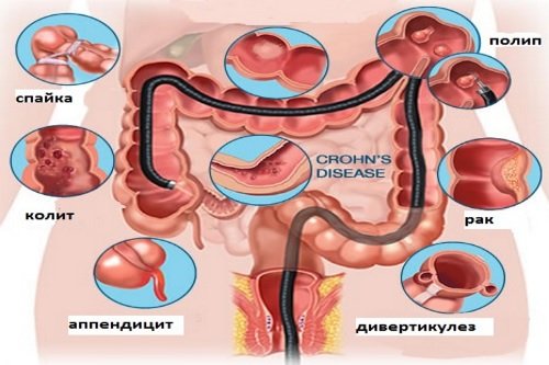
If during the examination the doctor suspects the patient has any intestinal pathology, he will prescribe a detailed examination. Colonoscopy has always been considered the gold standard.
But there are moments that do not allow its use (not all medical institutions have the necessary equipment and there are a number of contraindications to its implementation, including pregnancy, ulcerative colitis, some characteristics of the patient’s body, Crohn’s disease or the presence of a disease such as diverticulitis in remission) .
In such cases, instead of colonoscopy, other methods of examining the intestines may be used.
Hydrogen test
The procedure requires the patient to sit in one position for several hours. Every half hour, he must breathe into a device that calculates how much hydrogen is released within the small intestine by bacteria.
The fact is that microorganisms disrupt the absorption of fluid by the intestinal mucosa, as a result of which the patient suffers from bloating and diarrhea. In this case, carbohydrates are quickly decomposed into their component parts, and hydrogen is released outside during respiration.
Sigmoidoscopy and anoscopy
Sigmoidoscopy.
Examination of the intestines without colonoscopy using this method requires the use of a special rectoscope apparatus (a plastic device that has a depth scale and lighting). The procedure, similar to a colonoscopy, is prescribed in case of bleeding or sphincter pain.
A special tube is inserted into the rectum to a depth of no more than 35 cm and makes it possible to examine the sigmoid colon. During diagnosis, the doctor can comprehensively assess the condition of the mucous membranes, blood vessels, measure the diameter of the lumen, detect scars, cracks and polyps.
For better viewing, air pumping is used. Since sigmoidoscopy causes significant discomfort to a person, it is often performed under general anesthesia.
Anoscopy.
The procedure is very similar to sigmoidoscopy, but in this case, the tube is inserted no more than 12 cm. The anoscope can take tissue samples for analysis.
Capsule endoscopy
This technique is an excellent alternative to intestinal colonoscopy and is recommended to everyone who, for one reason or another, does not have the opportunity to undergo it. For this, a special mini camera equipped with a light source is used. It is covered with a shell on top.
The patient needs to swallow this capsule and put on a cuff in which a recording device is placed, which records the necessary indicators obtained directly from the capsule.
The convenience of this diagnostic method is that the patient does not need to be distracted from his daily activities.
Passing through the entire digestive tract, the tablet will take photographs and various measurements to identify possible diseases of the gastrointestinal tract. Many people believe that this is a worthy alternative to colonoscopy.
After 6-8 hours, the tablet with the camera will naturally leave the body, and the doctor will have all the information he needs. The only disadvantage of this procedure is the inability to take a tissue sample for analysis.
Ultrasound, MRI and CT
These types of diagnostics can also be used as an alternative method of colonoscopy:
- Ultrasound. Unfortunately, this method is not possible to qualitatively examine the patient. Ultrasound does not make it possible to determine oncological diseases at the initial stage of their development. Ultrasound examination checks for the presence of metastases in the case of intestinal cancer.
- MRI. Using magnetic resonance imaging, you can examine the intestines for the presence of large tumors and foreign objects. If you pre-administer the substance gadolinium, you can diagnose polyps.
- CT. When choosing a way to examine the intestines without a colonoscopy, sometimes they stop at computed tomography. However, it cannot give a complete picture in the case of cancerous tumors, since it is quite difficult to assess the condition of small tumors. At the same time, CT provides a clear image of the organ and does not damage the mucous membranes and skin.
Non-instrumental diagnostic methods
Gastroenterologists believe that if diseases are caused by nutritional disorders and do not have a serious etiology, then non-instrumental research options are allowed.
In such cases, palpation of the peritoneum, tapping, auscultation and visual inspection of the abdomen are performed.
Some diseases can be identified by hollowness or, conversely, swelling, the location of unpleasant sensations and their nature (dull, sharp, etc.).
In such cases, a history, urine and blood tests, liver tests and examination of the pancreas are sufficient to make a diagnosis. Proctologists, in turn, check the condition of the intestine using the anal-digital method, when the elasticity of its walls and mucous membranes is examined.
Source: https://proctologi.com/kishechnik/proverit-bez-kolonoskopii.html
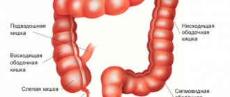
Instrumental diagnostic methods
Instrumental diagnostics is an examination of the intestines using specialized equipment. This group includes radiation research methods (x-ray, MRI, ultrasound) and endoscopic diagnostics (sigmoidoscopy, colonoscopy).
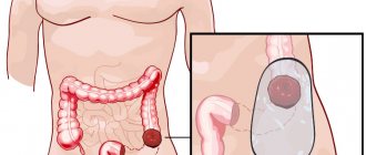
Colonoscopy
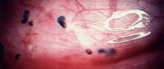
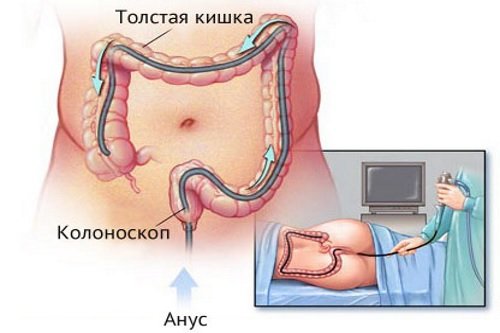
Colonoscopy is an examination of the large intestine using special endoscopic equipment that contains a camera and lighting elements. When the endoscope passes through the intestine, it “removes” the intestinal wall, and the resulting image is transmitted to the monitor. Thanks to this, the doctor can assess the nature of changes in the intestinal mucosa and detect pathological changes:
- tumors;
- cysts;
- inflammatory processes;
- polyps;
- areas of ulceration;
- local hemorrhages.
Some therapeutic manipulations can also be done directly during the diagnostic procedure. So, with the help of an endoscope, bleeding is stopped or polyps are excised. From suspicious areas of the intestine, you can take a biopsy - a fragment of the mucosal surface.
The biological material is sent for histological examination, where its cellular composition is determined.
We recommend reading:
Sigmoidoscopy and colonoscopy: what is the difference between the studies and which is better?
A biopsy is performed in patients with suspected malignancy. This technique allows you to accurately confirm the nature of the tumor and carry out differential diagnosis with benign changes in the mucosa.
X-ray examination
X-ray examination of the intestines is divided into main types - radiography and irrigoscopy. Plain radiography has little diagnostic value, since it cannot visualize intestinal loops. However, the image will show severe disturbances in the digestive system:
- places where fluid accumulates;
- the appearance of gas in the abdominal cavity (may indicate intestinal perforation);
- intestinal obstruction, which is characterized by the accumulation of the contents of the digestive system in one area and the lack of movement of food masses.
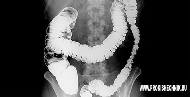
Irrigoscopy
Irrigoscopy is of greater value in diagnosis. The technique involves introducing a contrast agent into the patient’s gastrointestinal tract - a composition that is clearly visible on an x-ray. By the nature of the distribution of contrast in the digestive system and the speed of its passage through the stomach and intestines, one can judge the functional activity of the organ.
Irrigoscopy allows you to detect:
- pathological changes in the walls of organs (ulcers, tumors and polyps protruding into the intestinal lumen);
- narrowing of intestinal loops;
- the presence of accumulations of food masses (intestinal obstruction);
- inflammatory changes on the surface of the mucosa.
Irrigoscopy is an important component in diagnosing diseases of the gastrointestinal tract.
Ultrasound diagnostics
Ultrasound is an examination technique that is based on the reflection of ultrasound rays from the organs of the digestive system and their recording by a special sensor. The technique is completely safe for patients and can be used if there are contraindications to radiation exposure.
Using ultrasound, you can evaluate the size and shape of intestinal loops, the thickness of the mucosa, and the structure of the organ wall. On the screen you can detect signs of pathological changes (tumors, inflammatory processes). In addition, ultrasound is used to examine the liver and pancreas, with diseases of which it is necessary to differentiate disorders in the intestines.
CT scan
Computed tomography (CT) is a highly sensitive diagnostic technique that can even obtain layer-by-layer images of the abdominal organs.
Tomography allows you to identify signs of the following diseases:
- polyps and other benign neoplasms;
- malignant tumors;
- inflammatory diseases;
- intestinal bleeding.
The high accuracy of images makes CT an additional research method, which is used in complex diagnostic cases.
Colonoscopy
Colonoscopy of the intestines as diagnostics
Colonoscopy is widely used in proctological practice. It is an endoscopic method for diagnosing the entire large intestine and the beginning of the small intestine. Through it, diseases of the large intestine are diagnosed and the condition of the organ’s mucosa is assessed. To carry out the procedure, a special device is used - a colonoscope. Colonoscopy can reveal:
- benign and malignant tumors of the colon;
- nonspecific ulcerative colitis;
- Crohn's disease;
- colon polyps;
- intestinal obstruction;
- diverticula;
- bleeding.
If there are acute infectious processes in the body, then this procedure is contraindicated, as well as in case of peritonitis, blood diseases, heart and pulmonary failure. Severe forms of ischemic and ulcerative colitis are also contraindications for colonoscopy.
To obtain reliable data, it is necessary to prepare appropriately for this procedure. Proper preparation involves following these recommendations:
- Follow a diet that includes light meals for 2 days before the examination.
- Complete cleansing of the intestines from contents using special medications. If you have regular bowel movements, you can get by with an enema - before and the day before the examination.
- On the day of diagnosis and the day before, only liquid is taken as food, this can be tea, broth, water.
The procedure is performed without the use of general anesthesia. It is used if the patient is a child under 10 years of age. Local anesthesia is indicated for persons with increased pain. For examination, the patient must take a horizontal position. The examination is carried out by inserting a colonoscope into the anus.
Comparative table of intestinal diagnostic methods
| Diagnostic method | Advantages | Flaws |
| Colonoscopy |
|
|
| Radiography |
|
|
| Irrigoscopy |
|
|
| Ultrasound diagnostics |
|
|
| CT |
|
|
We recommend reading:
Checking the intestines without colonoscopy: alternative research methods
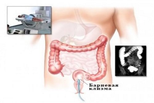
Irrigoscopy
Irrigoscopy of the intestine
Among the methods for diagnosing intestinal pathology, irrigoscopy occupies a special place. It is an examination using contrast-enhanced X-rays. This method is used primarily for examining the colon. After introducing a contrast agent into the patient’s intestines as it fills with it, overview and targeted images of the organ are taken. Barium sulfate diluted with water is most often used as a contrast agent. The procedure is painless, and despite its radiation exposure, it is safe, since such exposure is very insignificant.
This study shows the location of the intestine, its diameter and shape, and also evaluates the elasticity and distensibility of the intestinal wall. Irrigoscopy is the preferred method of examination when colonoscopy is not possible. It is also informative for signs of cancer of the colon. In addition, it is carried out if the following symptoms are present:
- bleeding from the intestines;
- purulent or mucous discharge from the intestines;
- pain in the anus and colon;
- chronic stool disorder.
For pathologies of cardiac activity, such as tachycardia and heart failure, and for some intestinal pathologies (diverticula and ulcerative colitis, perforation), this diagnostic procedure is contraindicated. Women who are pregnant cannot use this procedure either.
In what cases is it necessary to undergo examinations?
- severe abdominal pain;
- constant constipation;
- frequent diarrhea;
- the appearance of formations in the abdominal cavity;
- intestinal obstruction;
- change in the nature of feces, the appearance of blood and fat drops in it;
- itching and burning in the anal area.
In continuation of the topic, be sure to read:
- How to check the intestines for oncology?
- Intestinal ultrasound or colonoscopy: comparison of techniques and their information content
- MRI of the intestine and colonoscopy: which study is better?
- How is capsule endoscopy of the intestine performed?
- Preparation and performance of intestinal ultrasound
- Preparation and performance of computed tomography (CT) of the intestine
- X-ray of the intestines: the essence of the procedure, preparation and implementation
- Intestinal pneumatosis on ultrasound: what does the pathology mean and how is it treated?
- Irrigoscopy and colonoscopy: which test is better?
- Methods for examining the small intestine, their advantages and disadvantages