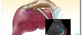
Ultrasound examination (ultrasound) is one of the types of complex non-invasive, practically safe examination of internal organs. Answering the question of which organs are checked during an ultrasound of the abdominal cavity, one should name:
- liver;
- gallbladder and its ducts;
- retroperitoneal space (together with the kidneys and adrenal glands);
- peritoneal cavity and vascular condition;
- pancreas.
In general, examination of the gastrointestinal tract using this technique is very effective. Let's find out who needs such a diagnosis and how to best prepare yourself for it.
- Indications for ultrasound
- Proper preparation Nutrition
- Medicines
- Purgation
- Smoking, chewing gum, lollipops, etc.
- Children
Indications for ultrasound
To identify the condition of internal organs, pathologies or consequences of injuries, an ultrasound procedure is prescribed. The main indications for the examination are:
- pain, groundless bitterness in the mouth, heaviness and other uncomfortable feelings in the stomach;
- frequent nausea;
- increased flatulence;
- temperature increase;
- suspected inflammation, infection, malignancy or functional disease.
Important! Experts recommend undergoing a comprehensive examination of the abdominal organs annually.

During the examination, you can determine the causes of pain and discomfort in the stomach, identify inflammation, cirrhosis, hepatitis, tumors and cysts, and the presence of stones in the gall bladder. The procedure is definitely prescribed after injuries, especially sports ones.
Ultrasound is a safe examination, as evidenced by the fact that it is periodically prescribed to pregnant women: the first, second and third are planned; in addition, cervicometry and doplerometry are done - types of ultrasound examination.
Ultrasound in the abdominal cavity: purpose
Purpose of abdominal examination
An ultrasound examination of the abdominal cavity must be performed if the patient has any suspicions regarding the health and performance of his internal organs.
Such an examination is necessarily carried out if the patient develops pathologies in the structure of the abdominal organs and identifies the factors of their occurrence.
Most often, ultrasound of the abdominal structures is performed for the following patient complaints:
- Activation of gas formation process
- The appearance of bitterness in the mouth
- Feeling of sharp pain in the abdominal area
- Feeling of heaviness in the right hypochondrium
- Perception of fullness after eating
- The emergence of heaviness
In addition to a person’s complaints about these symptoms, an ultrasound of the abdominal area should be performed if the following diseases are suspected:
- The appearance of various types of tumors
- The presence of various types of inflammation
- Manifestation of cholecystitis
- The occurrence of pancreatitis
- For hepatitis
- Suspicion of liver cirrhosis
Ultrasound is prescribed not only for adults, but also for children. Using this procedure, you can identify various pathologies and diseases that occur in acute or chronic forms. Timely ultrasound examination of organs located in the abdominal area allows not only to detect the appearance of changes in the structure of these organs, but also to establish the cause of their appearance, and also to prescribe urgent and correct treatment.
Preparation
Proper preparation for abdominal ultrasound
Before examining organs located in the abdominal cavity, you need to undergo a little preparation. It consists in meeting certain requirements, which will affect the final result of the study.
This examination is carried out on an empty stomach, so the patient should not eat anything in the morning. Even having dinner the evening before the procedure is not a big deal. Before undergoing an ultrasound examination of the kidneys, the patient is recommended to take about one and a half liters of fluid. This can be boiled water or juice.
This event allows you to fill the bladder and helps improve the performance of this examination.
Before performing an ultrasound of the abdominal organs, the patient should not undergo irrigoscopy and radiography for at least two days. If for some reason such examinations were carried out, then you need to inform the doctor about this so that he reschedules the ultrasound examination at a later date.
The reason for this is the use of barium during irrigoscopic and X-ray examinations, under the influence of which the results of ultrasound examination of the abdominal organs can be distorted.
Before the ultrasound procedure itself, it is recommended to adhere to a special diet, which should last about three days:
- It is based on cereal porridges, lean meats and fish, soft-boiled eggs, and low-fat cheeses.
- This diet excludes the intake of white breads, cabbage, apples, corn, dairy, beer and carbonated drinks, as well as sweets.
- You should avoid chewing gum, which promotes the swallowing of excess air.
- In this case, meals should be five times a day and in fractions.
- Smoking before the study is also not recommended.
Ultrasound procedure
Abdominal ultrasound procedure
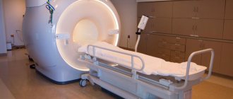
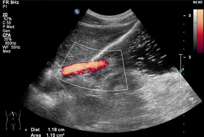
To conduct an ultrasound examination of the abdominal organs, special equipment is used, which works by producing high-frequency waves. This equipment produces sound pulses that are reflected from tissue structures that have different acoustic impedances.
The reflected waves are recorded by a special sensor. This produces not only a two-dimensional, but also a three-dimensional type of image of organs. Thanks to such devices, it is possible to record even the work of the abdominal organs, creating a three-dimensional image.
Features of abdominal ultrasound:
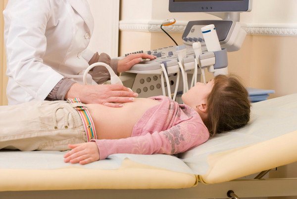
- When performing an ultrasound examination of the abdominal area, the patient does not feel pain, and no harm is caused to his body.
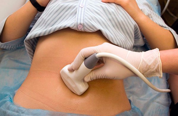
- The patient bares his body to the waist and lies sideways or with his back on the couch. The doctor must inform you about the adoption of a particular position.
- A thin layer of a special type of gel-like substance is applied to the surface of the patient’s skin, helping to fill air cavities to improve the penetration of ultrasonic waves.
- In some cases, the patient is asked to hold his breath for a while to better study the structure of the internal organs.
- The examination time on average lasts about thirty minutes.
- This procedure can be carried out not only in the morning, but also at lunchtime. This will not affect the results in any way.
Decoding the results
As soon as the attending physician receives the results of the study, he immediately begins drawing up a study protocol. This continues for several days. If any doubts arise about the results obtained, the patient may be prescribed a re-examination, which is the norm in many clinics.
Ultrasound examination provides the most accurate diagnosis of the condition of the internal organs of the human body. It is she who is the first to signal changes occurring in the body, which sometimes have a negative effect on the body.
Based on the results obtained from ultrasound in the abdominal cavity, the appearance of inflammation, changes in the size of organs and their displacements relative to each other are judged.
The cause of organ displacement is most often proliferating tissue, cysts, emerging neoplasms, expanding aortas with aneurysms, as well as processes leading to thickening of the walls of the gallbladder. The appearance of stones in the structure of the kidneys, dilation of the bile ducts, the appearance of solid formations in the internal structure of the gallbladder, as well as numerous types of damage to the abdominal organs are also detected during an ultrasound examination of the body.
Only experienced doctors can decipher the data from the obtained images and establish a diagnosis appropriate to the patient’s health condition.
More information about abdominal ultrasound can be found in the video.
Read: Main causes and signs of gallbladder disease
Modern equipment used for ultrasound examination allows one to obtain images of internal organs in fairly high quality, thanks to which doctors quickly determine the nature of changes that have appeared in the body and identify their causes. But sometimes the results obtained from ultrasound leave much to be desired, although they are carried out using highly professional equipment. The picture of ultrasound results can be distorted by the patient himself who behaves incorrectly during this procedure. To prevent this from happening, you need to listen and follow all the doctor’s commands, while maintaining a motionless position.
Too much weight of the patient due to severe obesity will somewhat interfere with obtaining a clear picture of the condition of the patient’s internal organs, due to the distortion of fat cells emitted by a special ultrasound device. Open types of wounds on the body can also interfere with obtaining an objective picture of the structural structure of the abdominal organs. In such cases, the patient is necessarily recommended to undergo additional diagnostics. To facilitate the doctor’s work when determining the diagnosis, the patient should take with him to the ultrasound the results of such procedures that were obtained earlier.
Proper preparation
Proper preparation for the abdominal ultrasound procedure is the key to obtaining objective results. The study can be damaged by the presence of air in the intestines, so the task of preparation is to eliminate it. The presence of a large percentage of subcutaneous fat is another obstacle to ultrasound. Therefore, a few days before the examination, it is important to organize proper preparation for an ultrasound of the abdominal organs.
Nutrition
3 days before the scheduled test you need to follow a special diet . Beans, any pastries, bread and confectionery products, milk, fruits and vegetables containing fiber, carbonated water and drinks are completely excluded from the diet.
It is also important to follow a special diet based on the consumption of fish and lean meat. Add baked apples and cereal porridge to your diet. Eat food in small portions, cook dishes in water without oil or steamed, and under no circumstances overeat.
Did you know? During the First World War, ultrasonic vibrations were used to determine the distance to the target and the location of enemy submarines.
Medicines
In the absence of any contraindications, the attending physician may prescribe medications. This applies to medications taken on a regular basis, and to medications prescribed immediately before the test for good digestion and reduction of gas formation. Thus, secretory function is improved by Festal and Mezim and some enterosorbents (Smecta, Enterosgel).
Important! Self-prescription of any medications is not recommended. Each individual drug is prescribed only in a specific case.

Purgation
Intestinal cleansing is an important preparatory step for patients suffering from various disorders of the intestinal tract. It is important that the intestines are completely empty before diagnosis . Experts recommend that patients with stool problems take a laxative (10-12 hours before the test). If after such manipulations the intestines still do not empty, a cleansing enema is performed.
Many people wonder: is it possible to drink water before an abdominal ultrasound to prepare for the examination? And the answer to this question is yes, if we talk about kidney examination. Carrying out such a diagnosis is possible only with a full bladder. To do this, drink up to 1.5 liters of liquid the day before (preferably still water).
Smoking, chewing gum, lollipops, etc.
Alcohol, as well as the use of chewing gum, candies and sweets, must be completely eliminated 2-3 days before the examination. In order not to distort the diagnostic results, you also need to refrain from taking no-shpa and aspirin. Separately, you should consider smoking, which causes stomach spasms.
This habit should be eliminated at least a few days before the ultrasound. Immediately before the examination you should absolutely not smoke in order to get the most reliable result.
Important! The last meal is taken 5-6 hours before the procedure. The study itself is carried out on an empty stomach.

How is the examination carried out?
The examination is carried out using high-quality diagnostic ultrasound equipment. The procedure takes 20 minutes. The technique is effective, accessible and painless, therefore it is recommended for both adults and children.
If you are preparing to conceive a child, you will be interested in knowing what fertility and ovulation are.
Children
When preparing a child for an abdominal ultrasound, you need to know what is included, namely good preparation. An important part of it is a diet that excludes foods in the list above. 6-8 hours should pass between the last meal and diagnosis, 3-4 hours for infants. When dressing your child, choose clothes that easily expose the stomach area.
Directly during the procedure, the child is placed on a couch, the skin is lubricated with a special conductive gel, and an apparatus with a sensor is easily moved over the surface of the stomach. This viscous gel is transparent, odorless, does not cause allergic reactions and conducts ultrasonic waves well. At the end of the session, the remaining gel is easily removed with napkins.
During the examination, the diagnostician can turn the child on his side to better view the organs on the monitor. Older children are often asked to take a deep breath and hold their breath to get the clearest image on the screen.
Adults
Traditionally, diagnostics are prescribed in the morning. According to doctors, this time is the most acceptable, since the patient has not yet become hungry and is not forced to endure “deprivation.” In some cases, diagnostics are prescribed in the afternoon, but then the patient needs to deny himself meals.
In some cases, unsweetened tea for breakfast is allowed. For research, special ultrasound devices are used - echotomoscopes. The procedure for adults is the same as for children.
Did you know? Today there is practically no organ that cannot be viewed using ultrasound. For example, ultrasound is used to determine brain damage and diseases.
How does the stomach ultrasound procedure take place?
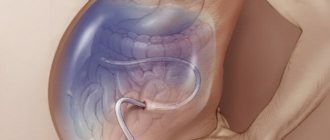
The doctor will ask you to lie down on the couch. The doctor will apply a water-based gel to the abdomen. Now there will be no air between the sensor and the skin and the image on the screen will be clear. The doctor will move a sensor over the skin and examine the condition of the gastrointestinal tract organs from different angles. Some time will pass and the doctor will ask you to drink water or juice through a tube.
On the monitor screen, the doctor will see how the liquid goes through the esophagus into the stomach. The liquid will help to perfectly visualize the various departments. This examination will end and the ultrasound specialist will immediately give you a conclusion. The doctor will read it and sign it, stamping it. The results will be recorded on a computer disk.
Thanks to the recording, your attending physician will look at the state of the gastrointestinal tract now and will be able to trace all pathological changes that may occur in the future.
Ultrasound is not recommended after such studies:
- FGDS;
- Colonoscopy;
- Irrigoscopy.
- Gastrography.
An ultrasound examination of the stomach is an effective diagnostic method, but it does not replace X-ray or endoscopic examination. Thanks to ultrasound, the contours of the stomach and the condition of its walls are visible. After a while, it will be possible to do it again and find out if there is any regression, if the pathology is growing?
Diagnostic results
What does an abdominal ultrasound show? First of all, the diagnostician evaluates:
- size, position, structure and shape of the liver;
- the presence of inclusions such as stones;
- general condition of the gallbladder, its ducts and vessels of the cavity;
- pancreas, spleen and kidneys.

Based on the results of the echogram obtained using an ultrasound machine, the doctor makes a conclusion about the general condition of the upper abdominal cavity. The subject receives a protocol form with recorded normative indicators and specific data.
In some cases, the results obtained are unreliable if the patient has extreme obesity or increased flatulence due to improper preparation. In such cases, additional examination is prescribed.
Health Ultrasound examination