This is one of the most informative diagnostic methods, which is suitable for identifying most digestive disorders.
Essence of the method
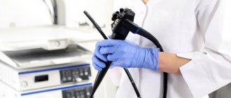
Endoscopy is performed using a gastroscope. A gastroscope is a device consisting of an elastic tube with a complex optical system and a visualization device. The tube is sequentially inserted into the pharynx, esophagus, stomach and the initial part of the small intestine. Thanks to fiber optics and digital equipment, the doctor can see structural changes in the mucous membrane of the digestive tract and evaluate its functional features.
Information. The abbreviations FEGDS and EGDS are synonyms. The term fibroesophagogastroduodenoscopy (FEGDS) emphasizes the important role of fiber optics in the operation of the device (from the Latin fibra - fiber).
You can learn about the operating features of modern gastroscopes from the video.
When using special equipment, additional diagnostics become possible: microscopy of the mucosa after taking biopsy material and intragastric pH-metry - determining the level of acidity of gastric juice.
Method capabilities
Visual methods allow the diagnostician to directly see in real time the condition of many internal human organs. First of all, this applies to most parts of the food tube, that is, the pharynx, the walls of the esophagus, the stomach cavity, the small and large intestines, as well as the bile ducts and the gallbladder itself are subject to inspection. Thus, it is possible to directly examine the condition of these organs, identify erosions, inflammatory or ulcerative-necrotic changes, tumor processes, and also evaluate the degree of effectiveness of treatment using the resulting morphological picture or be guided by them during surgery. However, the capabilities of these methods are not limited to this, because with the help of probes you can also deliver medicine precisely, take portions of gastric or bile juice, as well as biopsies from various parts for analysis. Thus, the doctor will be able to get a complete picture of what is happening in the body, make a correct diagnosis in a timely manner and correctly prescribe treatment.
Indications for use
Esophagogastroduodenoscopy is prescribed for suspected diseases and functional disorders of the upper gastrointestinal tract (GIT):
- esophagitis;
- gastritis;
- duodenitis;
- neoplasms;
- anomalies in the structure of organs;
- peptic ulcer;
- erosion;
- foreign bodies;
- burns and injuries of the esophagus, stomach;
- biliary or gastroesophageal reflux;
- diverticula;
- stenosis of the esophagus, cardia of the stomach.
When pathology is detected, EGDS is sometimes repeated over time to assess the quality of treatment or the rate of disease progression.
FEGDS is prescribed not only for diagnostic, but also for therapeutic purposes:
- to stop bleeding;
- to remove polyps;
- if necessary, administer the drug directly into the gastric cavity;
- for the purpose of removing a foreign body.
Indications and contraindications for examination
Having understood what FGDS is and how it is done, it is worth studying the main indications for the procedure. Indications are divided into planned and urgent. Planned ones include:
- heartburn;
- feeling of nausea, vomiting;
- bloating;
- lack of appetite;
- rapid weight loss for no apparent reason;
- swallowing disorder;
- frequent pain in the abdomen.
Among the urgent ones there are:
- removal of foreign bodies;
- stopping gastrointestinal bleeding;
- assumptions about acute diseases of the duodenum and stomach.
What does FGD of the stomach show:
- gastritis;
- varicose veins of the esophagus;
- anemia of unknown etiology;
- peptic ulcer;
- tumor;
- complete or partial obstruction of the esophagus;
- esophagitis;
- GERD;
- decrease in the lumen of the duodenum;
- protrusion of the walls of the esophagus;
- DGR.
Contraindications
Most contraindications to gastroscopy are relative. The question of the possibility of conducting an examination is decided in each case individually. Typically, EGDS is prohibited when:
- severe cardiac and respiratory failure;
- low blood clotting rates;
- mental disorders in the acute stage;
- aortic aneurysm;
- mediastinitis – inflammation of the mediastinal organs;
- serious consequences of stroke (including swallowing disorders).
Absolute and relative contraindications to gastroscopy
There are no absolute contraindications to gastroscopy, that is, this study, if emergency diagnosis is necessary, can be carried out even in the presence of relative contraindications, but after preliminary preparation of the patient.
Relative contraindications to gastroscopy, depending on the presence of a particular pathology, can be divided into several groups:
- cardiovascular;
- hematological;
- musculoskeletal;
- metabolic and endocrine;
- neurological.
Preparation
To obtain the most informative results, proper preparation for FEGDS is important:
- During the week before the procedure, it is necessary to do all the tests and instrumental examinations prescribed by the doctor (blood test, urine test, ECG, etc.). This will help identify possible contraindications.
- 72 hours before the procedure, it is recommended to avoid eating irritating and too heavy foods. In particular, it is better to exclude marinades, smoked foods, overly spicy and salty foods, alcohol, and soda from the diet.
- It is recommended to stop eating approximately 10 hours before FEGDS, so that it has time to evacuate from the stomach.
- You can't smoke or drink 3 hours before. In the morning, the only drinks allowed are still mineral water.
- As a rule, before an endoscopic procedure, it is recommended to discontinue medications that reduce blood clotting (antipyretic, anti-inflammatory, painkillers) and suppress gastric secretion (omez, Nexium, ranitidine).
The use of any medications before manipulation is usually not necessary.
Note. Both prescription and withdrawal of medications are carried out only after consultation with the doctor.
Advantages of endoscopy
Esophagogastroduodenoscopy (EGDS) is one of the probing methods that allows you to examine the esophagus, stomach and duodenum using a microcamera. Of course, there is another method, non-invasive - x-ray examination after taking a barium suspension in the form of porridge to contrast the mucous membrane. However, this method has rather limited capabilities and allows you to see only chemical burns, tumor processes, ulcers and erosions in the image. While an EGD examination can also reveal bleeding, take biopsies for microscopic analysis and examine the organs in more detail. This technology also allows you to assess the condition of the departments after surgery, namely healing, integrity of sutures, bruises, remove foreign objects, and diagnose varicose veins of organs.
Technique
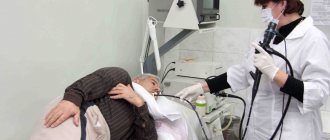
To conduct FEGDS, the patient is placed on his left side and premedicated: an anesthetic is applied to the pharyngeal mucosa, which inhibits the gag reflex, and sometimes sedatives are administered.
Peculiarities. Young children and adults are allowed to undergo general anesthesia or medicated sleep before endoscopy.
Next, a special mouthpiece is placed in the mouth, which prevents the teeth from closing and damaging the gastroscope.
As the gastroscope tube passes through the pharynx, the patient is instructed to actively swallow it. In the future, if possible, you need to remain calm and relaxed. The calmer your behavior is, the faster the doctor will receive all the necessary information.
The results are usually interpreted immediately. Thickening or thinning of the mucosa is considered a sign of gastroduodenitis. A cancerous tumor often looks like an uneven growth in the stomach wall. Benign neoplasms have a regular shape and clear contours. An ulcer is a defect in the mucosa, sometimes with signs of bleeding. The presence of reflux is determined by the backflow of intestinal contents into the stomach or from the stomach into the esophagus.
The essence of EGDS
Many patients who have received a referral from their attending physician for this examination often ask the question: “EGD, what is it?” The abbreviation usually frightens patients and even confuses those who have never encountered visual diagnostics before. So let's try to figure out how it works. Endoscopy of the stomach or other part of the gastrointestinal tract is carried out using a special probe - a flexible, controlled tube consisting of an elastic material and additionally having fiber optics (a kind of camera and light bulb) and manual control technology. This allows you not only to lower it along the gastrointestinal tract, but also to turn it to the sides or down to get a complete visual picture. After the examination is completed, the probe is carefully removed so as not to create iatrogenic mechanical damage to the internal membranes of the organs.
Possible complications
Dangerous complications of the procedure include bleeding from the gastrointestinal tract during FEGDS with biopsy and perforation of the organ wall with a gastroscope with subsequent development of peritonitis. Symptoms of these conditions usually appear during or immediately after the procedure. They require emergency medical attention.
For a couple of days after endoscopy, the patient may experience discomfort in the pharynx due to damage to the inner layer of the epithelium. This discomfort usually goes away spontaneously, without outside intervention.
Delayed and rare complications include infectious inflammation. Bacteria and other pathogens enter through improperly processed medical instruments. This condition is accompanied by an increase in all symptoms: pain in the epigastrium and behind the sternum, nausea, vomiting, heartburn. Therefore, you need to treat your condition carefully; if your health worsens, be sure to inform your doctor.
Preparing for a stomach examination
When diagnosing, it is quite important to correctly assess the mucous membrane of the gastrointestinal tract, so the process of preparing for the study should be completed.
Gastroscopy is performed only on an empty stomach. Experts recommend that the last meal be taken at least 6-8 hours before diagnosis. Usually the procedure is done in the morning, so in the morning the patient should also not consume anything liquid or food.
If the patient wears removable dentures, they must be removed before diagnosis. A few hours before the procedure, you should stop smoking. If the study is performed under anesthesia, then it is advisable to extend the fasting period to 10-12 hours.
Precautions during the procedure
Since most patients, especially children, are afraid of this study, the doctor must explain to each of them before endoscopy that this, although unpleasant, is a very necessary method for maintaining their health. He should be told that there are probes of different diameters, it takes a little time (literally 20-30 minutes), and immediately before its introduction, the oral cavity is anesthetized using novocaine in the form of an aerosol. This is also necessary to suppress the gag reflex, since the probe will irritate the uvula and pharyngeal baroreceptors. Therefore, if the patient feels a bitter taste in the mouth or slight swelling of the tongue, this is an absolutely normal reaction to the drug. In addition, after anesthesia, a mouthpiece is inserted - a sterile plastic device to protect the lips and teeth of the subject during insertion of the probe into the gastrointestinal tract.
Esophagogastroduodenoscopy
What is zophagogastroduodenoscopy (EGD)?
This is a safe and highly informative method for examining the esophagus, stomach and duodenum using an endoscope. Endoscopy allows you to visually assess the condition of the mucous membrane of the upper gastrointestinal tract and, if pathology is detected, take material for histological examination (biopsy).
To whom and why can EGDS be recommended:
For diagnostic purposes for:
- dyspepsia (nausea, belching, vomiting);
- pain in the upper abdomen;
- heartburn (feelings of acid or bitterness, burning in the throat or chest);
- bleeding (vomiting blood or blood in the stool);
- dysphagia - difficulty swallowing (food or liquid gets stuck in the esophagus);
- pathology identified on an x-ray of the upper gastrointestinal tract.
Additionally, during the study can be performed
- biopsy (for subsequent histological examination);
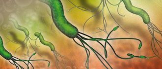
- determination of Helicobacter pylory
in a biopsy of the gastric mucosa.
For medicinal purposes:
- removal of stomach polyps;
- removal of foreign bodies;
- endoprosthetics (stenting) of the esophagus, stomach in case of tumor lesions;
- stopping bleeding from varicose veins of the esophagus and stomach (ligation), gastric and duodenal ulcers (combined hemostasis).
Advantages of endoscopy
Modern endoscopic systems allow examination in different light modes (trimodal edoscopy), obtain a comprehensive assessment of the characteristics of the mucous membrane, and are also a convenient tool for endoscopic screening of pretumor pathology and cancer.
How to properly prepare for research:
- Endoscopy is performed in the first half of the day, on an empty stomach.
- The last meal is a light dinner the day before no later than 19:00.
- It is possible to conduct the study in the 2nd half of the day; a light breakfast is allowed.
- We recommend taking with you the data from previous studies (gastroscopy, X-ray, CT, ultrasound).
How the research is carried out
The examination is carried out with the patient lying on his left side. A mouthpiece is placed between the teeth to prevent damage to the endoscope. Under visual control, the endoscope is passed into the esophagus and then, moving and turning it, the doctor examines the inner surface of the esophagus, then all parts of the stomach and duodenum. The enlarged image is displayed on a monitor, so the doctor can see minimal changes in the tissues. Through the channel of the endoscope, biopsies (small pieces of tissue) can be taken, fluid and air can be introduced and removed. The biopsy is painless.
During the examination, video recording is made from the endoscope camera, which the patient receives on a digital medium along with the conclusion.
Anesthesia
During endoscopy, the patient may experience discomfort when filling the stomach with air. To eliminate discomfort, the examination is performed under anesthesia: the anesthesiologist, using non-narcotic drugs, puts the patient into a short medicated sleep. The patient easily wakes up after the procedure and can begin daily activities within 2 hours.
Algorithm for the procedure
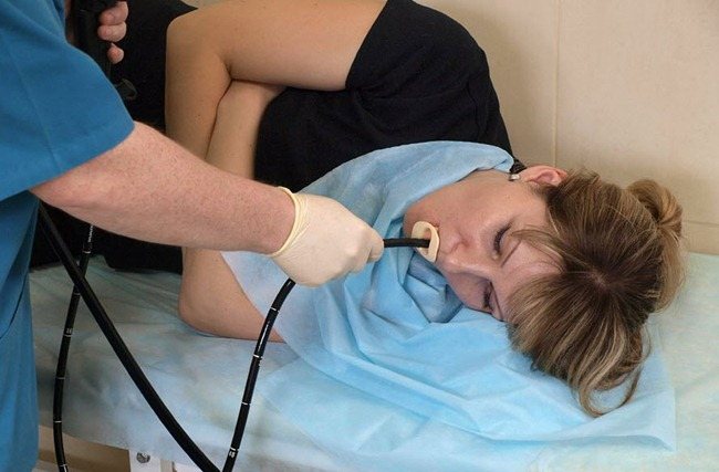
Gastroscopy is performed in several ways. The classic one is based on the introduction of an endoscope after local anesthesia of the pharynx. For emotionally labile patients, children or those with a strong gag reflex, transnasal gastroscopy or short-term anesthesia is performed. Inserting a probe through the nose allows additional examination of the vocal cords. Before manipulation, the specialist must configure the device and make sure it is working properly.
Traditional gastroscopy method
Gastroscopy of the esophagus and stomach is done in the morning. The patient's throat is irrigated with lidocaine aerosol, which will make it easier to tolerate the insertion of the endoscope and reduce the gag reflex. The patient is placed on his left side and asked to bend his knees. A ring-type mouthpiece is inserted into the mouth so as not to damage the endoscope with teeth, and a saliva ejector is used. When the muscles of the pharynx become numb, a tube is inserted through the mouth. It is recommended to breathe through the nose. Uncomfortable sensations disappear if you follow the advice and recommendations of medical personnel. The doctor checks the walls of the esophagus and stomach for 20-25 minutes, identifies pathological changes, and examines the tissues adjacent to them. If necessary, takes biopsy material and gastric secretions for analysis, and applies the medicine to the damaged surface. Surgery may also be performed to remove polyps or cauterize bleeding vessels.
A repeat gastroscopy can be performed a month after the previous one. That's how long it's valid.
Gastroendoscopy in a state of medicated sleep
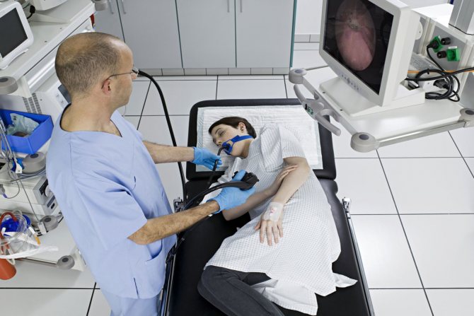
Esophagogastroduodenoscopy is an unpleasant but informative procedure. Often the patient refuses it due to fear of painful sensations. In this case, as well as for children, other diagnostic methods are offered. You can give intravenous anesthesia, which will allow you to carry out the procedure painlessly, or use a special capsule. It is swallowed and “collects” information about the state of the digestive tract. The state of medicated sleep is a safe and complete replacement for classical gastroscopy. The algorithms for the actions of the patient and the doctor are identical to those described above.
