When palpating the stomach at first, the tumor is usually not palpable. In order to feel it, it is necessary, on the one hand, to have favorable conditions for palpation, and on the other hand, for the developing tumor to reach a certain size (from the “plum”). Palpation of the tumor is often hampered by the stomach being full of food and some abdominal tension; To improve palpation conditions, it is necessary to conduct the study on an empty stomach and after thorough gastric lavage. It is useful for the patient to cleanse the intestines with a laxative beforehand. The study must be carried out with the patient standing and lying down, since small tumors of the lesser curvature sometimes become accessible to palpation only in an upright position, when they emerge due to prolapse of the stomach from the hypochondrium or from under the liver.
Probing is carried out systematically. A necessary prerequisite for a correct examination of the stomach is to clarify the topographic relationships of the abdominal organs using the method of deep sliding palpation, including the stomach and its relationship to neighboring organs, and the entire area of the stomach is examined and in the end it is still possible to palpate a tumor somewhere, or in the shape of a roller, or a scallop. It should be noted that in some cases, the palpable tumor of the pylorus can be extremely mobile and can be easily displaced both to the right and left hypochondrium, sometimes to the pubis; Thus, the significant mobility of the tumor and the location of the tumor outside the epigastric region do not exclude its belonging to the stomach.
During a methodical examination of the stomach, it is often possible (in 25% of healthy people) to palpate the pylorus, which during contraction reaches great density, however, unlike the tumor of this part of the stomach, it is completely smooth, and during relaxation it loses its distinct contours.
The palpable tumor, depending on its structure, has a different consistency: sometimes it is hard, uneven or lumpy in scirra, sometimes it is relatively soft, giving a vague feeling of resistance in the area of the stomach or “stomach filled with cotton wool.” Palpation of a cancerous tumor is usually painless or slightly painful if there is no invasion of the peritoneum; An inflammatory tumor in the area of a stomach ulcer is always painful when palpated. Palpation of large stomach tumors is much easier. A very characteristic palpation picture is given by infiltrating scirrhus. In these cases, the entire small, high-lying stomach with dense and often completely smooth walls is palpated, which sometimes appears as a sausage-shaped tumor lying in the epigastric region.
Having palpated the tumor in the stomach area, you also need to establish that it actually comes from the stomach. Tumors of neighboring organs may be located in the same area where the stomach should anatomically be located. It is necessary to distinguish tumors arising from the left lobe of the liver, omentum, transverse colon, tumor of the spleen or pancreas. It is especially difficult to determine the source organ for the tumor if adhesions develop around it. It is typical for a stomach tumor that it is located in the zone of the tympanic sound of the stomach, there is respiratory and palpation mobility, and if the tumor is localized on the posterior wall, there is a splashing noise above it, but when the stomach is full, it is difficult to palpate. The difference in percussion sound allows one to distinguish a stomach tumor from tumors of the left lobe of the liver. Sometimes, when palpating above the stomach tumor, the edge of the liver is felt. A tumor of the fundus of the stomach can sometimes resemble an enlarged spleen, but the spread of the dull sound zone from the tumor upward to the chest, the remaining dull sound of the tumor when the stomach is inflated, as well as the tendency of the tumor to go into the left hypochondrium speak in favor of a spleen tumor.
Pancreatic tumors are almost immobile, easily transmit the pulsation of the aorta and disappear when the stomach is distended. With tumors of the pancreas, disturbances in its functions (steatorrhea, glycosuria) may be observed. Tumors of the transverse colon have a cylindrical shape and in most cases they have symptoms of intestinal stenosis.
Careful palpation of the liver is necessary both in case of suspicion and in case of an established diagnosis of gastric cancer. Detection of metastatic cancerous liver nodes makes surgical treatment useless. The liver is the organ most often affected by metastases in gastric cancer. Cancerous liver nodes usually grow very quickly and there is a significant increase in liver size. When palpated, an enlarged, lumpy, very dense, painless liver is determined. The edge of the liver is usually uneven; one or more hard nodes surrounded by dense liver tissue can be felt near the edge; a single node (or several) can be palpated on the surface of the left or right lobe of the liver. The discovery of an enlarged, lumpy liver does not necessarily indicate cancer metastases to the liver; such a liver may be due to echinococcosis, primary cancer, syphilis or cirrhosis; Detection of the growth of a particular node is of decisive importance, and in addition, a differential diagnosis of other liver lesions is necessary (history, functional tests). Cases of the development of cancerous nodes in the liver in the absence of stomach symptoms are not very rare, and the diagnosis of the primary tumor is established only at autopsy.
Structure of the tumor and its types
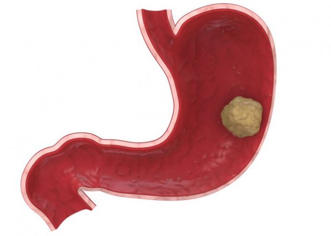
A lump in the stomach is a walled body filled with liquid or gas. The size and composition of the cyst directly depend on the cause of its formation. The size of the cyst can vary from 1 to 200 millimeters.
The location of the seal is the pyloric area. It is because of this that it can impede the movement of food through the gastrointestinal tract.
The cyst can be of two types:
- false – without epithelial covering layer;
- true, containing in the structure a cell cover responsible for strengthening the walls of the neoplasm.
The most often diagnosed is a true cyst, the occurrence of which is provoked by an anomaly that arose during intrauterine development.
The main causes of lumps in the stomach
The systematization of cysts has a rather complex structure. This is due to the diversity of their shapes and sizes. Despite this, the causes of such pathologies have long been well studied:
- a congenital factor caused by a violation of the development of the gastrointestinal tract;
- diseases associated with inflammatory processes in the gastrointestinal tract;
- mechanical effects on the internal walls of the main digestive organ;
- burns of the gastric mucosa resulting from prolonged consumption of alcohol-containing drinks;
- harmful effects of various microorganisms.
The most common is the first reason, the risk of which cannot be excluded. Other factors that contribute to the formation of lumps in the stomach can be eliminated using basic preventive measures, the main one of which is a healthy lifestyle.
Factors associated with the occurrence of lumps in the stomach area
Any disease of the gastrointestinal tract entails a lot of problems that cause a decrease in quality of life. As a rule, all gastrointestinal diseases are related to each other, and one can develop into another, for example, gastritis into an ulcer, etc. A cyst, which can arise as a result of the following diseases, is no exception:
- Ménétrier's disease is a systematic disorder of the digestive system associated with the proliferation of the mucous membrane of the main digestive organ. This disease is most often diagnosed in middle-aged men. The causes are not fully understood;
- Zollinger-Elisson syndrome, characterized by excessive production of hormones, which in turn leads to blockage of the glands responsible for the production of digestive juice;
- herpes, which causes damage to gastrointestinal tissue;
- chronic syphilis, which causes damage to the mucous membranes throughout the body, including the stomach;
- pneumatosis, uncontrolled flow, which causes the formation of cysts filled with gas. This condition is typical for people who move little;
Timely treatment of gastrointestinal diseases allows the formation of tumors in the stomach area to be avoided, with the only exception being congenital pathologies. Thus, if you experience symptoms indicating stomach disease, you should consult a doctor.

Stomach tumor: signs, symptoms and diagnosis
A stomach tumor is a complex disease that poses a threat to human life.
It is the appearance of formations on the gastric walls, which can be benign or malignant.
Modern medicine and advanced equipment make this disease curable. The main thing is to promptly notice its signs, carry out diagnosis and proper treatment.
Benign formations, their signs and symptoms
Approximately 5-10% of all neoplasms on the gastric wall are benign tumors, and 90-95% are malignant. The first type includes formations that grow and develop rather slowly.
However, they are characterized by a favorable prognosis. However, if they are not identified in a timely manner and treatment is not started, they can develop into dangerous neoplasms.
Therefore, it is important to monitor your health and identify the first symptoms in time.
Different types of benign stomach tumors have different signs and symptoms. In most cases, such ailments practically do not manifest themselves at all. They can be identified during endoscopic examination.
When asking the question of how to indicate the degree of the disease, you should understand what types of tumors are found. The following types of benign diseases are distinguished:
- polyps;
- leiomyomas;
- lipoma;
- neuroma;
- angioma;
- fibroma.
The first type is most common. It can be single or multiple. How to identify this species? In this case, the following ailments are observed:
- nagging or aching pain that occurs in the epigastric region (they occur after eating or 1-3 hours after eating);
- belching after eating air or food;
- the appearance of nausea or vomiting reactions;
- discomfort in the sternum and burning;
- heartburn;
- frequent weakness, dizziness, loss of strength, rapid fatigue (this may be caused by hidden bleeding from the polyp).
The second type of neoplasm is leiomyoma. This tumor in the stomach does not have any symptoms. Signs of this disease may occur when part of the tumor dies or an ulcer appears on it. This leads to hidden bleeding in the stomach and the appearance of the following symptomatic manifestations:
- dizziness;
- frequent weakness and loss of strength;
- anemia;
- sudden weight loss.
The exact cause of the appearance of these benign formations has not yet been identified. However, most often the following factors lead to them: chronic gastritis, bad habits, poor environment, infection, decreased immunity, heredity.
Dangerous tumor: symptoms
Malignant neoplasms differ from benign ones in that their cells already differ from the type of cells of the organ on which the tumor arose. Such formations have partially or completely lost the ability to differentiate. They are a serious threat to human life. Malignant tumors appear especially often in men aged 50-70 years.
Depending on the stages of the disease (early or late), its symptoms also differ. Thus, an early symptom of a stomach tumor can be identified by the following ailments:
- loss of appetite;
- sudden weight loss;
- feeling of a full stomach;
- the occurrence of pain, as well as a feeling of heaviness felt in the upper abdomen (this is especially pronounced after eating);
- the appearance of a feeling of disgust for certain foods (for example, meat).
At a late stage, a malignant tumor of the stomach has completely different symptoms. At this stage, intoxication of the body occurs. It is called tumor or cancer. How to determine this stage? In this case, you can notice the following first signs of a stomach tumor:
- depressive states, general weakness of the body, lack of a sense of joy, fatigue, decreased or loss of interest in work and work;
- frequent headaches, slow reactions, dizziness, poor sleep at night, desire to sleep during the day;
- severe loss of appetite (can lead to anorexia or cachexia);
- pale, blue or yellowing of the skin;
- causeless increase in temperature;
- dry mouth, eyes and nose;
- excessive sweating (especially at night);
- deterioration of immunity and resistance to various diseases;
- anemia;
- causeless nausea and vomiting reactions;
- sudden abdominal pain, heaviness in the gastric region (especially severe after eating);
- vomiting of foods eaten the day before (accompanied by an unpleasant rotten odor);
- enlarged lymph nodes located in the neck;
- the presence of blood discharge in the stool.
Diagnosis of the disease
If you notice any symptoms of the disease, you should immediately consult a doctor. It will qualitatively and accurately identify a stomach tumor; diagnosis will be carried out using various methods. During this process, all problems and malfunctions in the body will be identified. This will help avoid serious health problems, and in some cases even save lives.
One of the reliable and advanced institutions where you can diagnose the gastrointestinal tract is the Road Clinical Hospital at the station. Bitter". It has the latest equipment that can give the most accurate results. Thus, the device with which the research is carried out is a special endoscope Exera III. It is manufactured by Olympus.
How to diagnose a disease using special equipment? Having decided to check the condition of your gastrointestinal tract using this equipment, you can be confident in the quality and accuracy of the data obtained.
In case of stomach problems, a procedure called gastroscopy, which is an upper endoscopy, is performed. During this analysis, the stomach, esophagus and duodenum are examined.
The essence of gastroscopy is as follows. A special device, which is a soft tube with a tiny video camera attached to it, is inserted through the mouth into the human digestive system.
As a result, an image of the mucous membrane of the diagnosed organs is displayed on the monitor, and an endoscopic picture appears.
If necessary, this image can be enlarged many times, which makes it possible to examine the surface under study in detail.
Diagnosis of a stomach tumor includes not only examination using special equipment, but also the study of various important information about the patient:
- analysis of complaints and symptoms (identification of all ailments that torment the patient);
- analysis of the patient’s life (presence of any gastrointestinal diseases, bad habits);
- family analysis (heredity and predisposition to disease are revealed);
- visual examination of the patient (study of the appearance of the skin, mucous membranes);
- blood test (detects anemia);
- stool test (presence or absence of blood in stool).
It is after identifying the disease that it is worth taking time to study how the tumor is treated. Particular attention to preventive diagnostics should be given to people over 50 years of age. After this milestone, the likelihood of tumor growth is greater than at an early age.
Even if there are no complaints or signs, you need to set aside time for the examination. This will help avoid serious problems in the future. Timely identification of problems will make it possible to begin high-quality and effective treatment, thereby preventing the development of benign formations into malignant ones.
Source: https://www.dkb-nnov.ru/opuxol-zheludka/
Symptoms characteristic of the appearance of seals in the stomach
As a rule, this type of pathology develops sluggishly without pronounced symptoms. Most often, a cyst is diagnosed only after it has grown to a large size and causes significant discomfort.
The main symptoms of its presence are:
- tingling pain in the stomach area;
- rapid weight loss;
- lack of appetite;
- nausea turning into vomiting;
- diarrhea;
- swelling in the arms, legs and face;
- heartburn and flatulence.
One of the above symptoms is not a reason to panic, but if they are combined and appear regularly, you should immediately consult a doctor.
How is intestinal palpation performed?
Intestinal palpation involves two types of palpation of the abdominal cavity: superficial and deep.
Superficial palpation is always carried out first. After it, the doctor conducts a more in-depth study of the intestines and its specific parts.
If the patient has painful areas, an important rule that the doctor follows is the following: in no case should palpation begin from the place that hurts. Usually the doctor starts from the opposite part of the abdomen.
Most often, palpation begins from the left iliac region and involves feeling the intestines in a circle and counterclockwise.
Video about the intestinal palpation technique:
Surface method
With the superficial palpation method, the doctor needs to relax the patient as much as possible. For this, the patient is placed in a horizontal position with his knees slightly bent. This way the abdominal muscles relax as much as possible.
If the patient is still too tense, the doctor can distract him from the procedure by forcing him to perform breathing exercises.
The palpation occurs very smoothly and accurately. The area that hurts is palpated last, since if you start the procedure from the painful area, the muscles of the anterior abdominal wall will tense, which will not allow a full examination.
Deep
A deep type of palpation is performed to diagnose serious changes in the structure of the intestine. The most important condition for deep palpation is the doctor’s clear knowledge of the projection of the internal organs onto the anterior abdominal wall.
For diagnostic accuracy, when performing deep palpation, the doctor feels not only the intestines, but also other organs of the abdominal cavity.
During deep palpation, the patient must breathe deeply, evenly and measuredly through the mouth. In this case, breathing should be diaphragmatic. To facilitate the procedure, the doctor artificially creates folds of skin on the patient’s abdomen and then moves the palm into the required position.
When palpating the intestines, the doctor always follows the following sequence of palpating the organs:
- sigmoid colon;
- cecum;
- ascending and descending;
- transverse colon.
During deep palpation, the doctor must determine the diameter, nature of mobility, rumbling and painful areas of all parts of the intestine.
Diagnostic methods
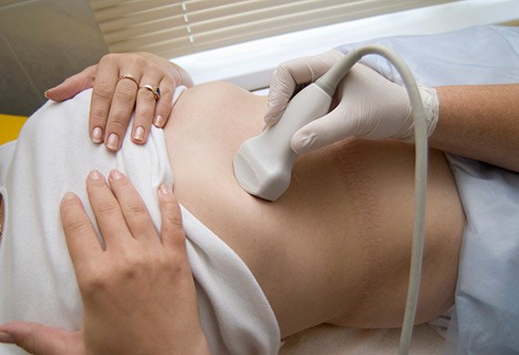
If a gastroenterologist suspects the presence of a cyst in the stomach, hardware tests are prescribed.
Diagnosis of lumps in the stomach can be carried out in the following ways:
- ultrasonography;
- gastroscopy;
- biopsy.
As a rule, at the initial stage of diagnosis, an ultrasound examination is performed, the result of which indicates the need for other procedures.
Can a lump appear under the skin on your stomach?
- Types of bumps on the stomach
- Causes
- Treatment
Any lump, including a lump under the skin on the abdomen, should not be ignored, as this formation may be a symptom of the development of pathology. These are mainly dermatological diseases that can be treated, but there is some possibility that disorders of a different nature may develop.
Types of bumps on the stomach
Formations under the skin may differ in origin, possible consequences for the body, as well as methods of treatment. Seals are also divided into benign and malignant, which rarely, but still, can form on the abdomen. If we consider the main types of cones, the following groups are distinguished:
- Atheroma. It is a benign type, formed from fat cells due to blockage of the glands. It feels dense, with clear boundaries and painless. Only in case of inflammation and suppuration does it cause discomfort and can open on its own. It is removed by surgical methods; conservative treatment for atheroma does not give positive dynamics.
- Cysts. At the initial stage, the formation does not have any special symptoms, but over time, compaction is noted, and this lump under the skin on the abdomen hurts. The sensation can be pulling, and as the size of the cyst increases, the pain increases. The formation rarely degenerates into a malignant tumor, consists of connective, adipose tissue, and has a cavity in which biological fluid accumulates. Treat exclusively promptly.
- Pimples. One of the harmless types of bumps that can form on the stomach. If you do not make efforts to squeeze out, but carry out the correct treatment, you will get rid of the problem area relatively quickly and without scars. Usually pus accumulates in pimples and if opened independently, there is a possibility of infection spreading to other tissues.
- Lipoma. Often called a wen, it forms in the subcutaneous layers, can have different sizes, and grow into other tissues. The compaction at the initial stages is soft, as it develops it can become denser, and practically does not degenerate into cancer mutation cells. The lump does not lead to pain; discomfort begins to appear when neighboring tissues are pressed, especially when nerve endings are pinched. If there is a need for this, it is recommended to remove the fatty tissue in a medical institution; opening it yourself can aggravate the situation, for example, the tissue becomes infected.
Remember!
In addition to benign neoplasms, some of which are listed above, there is a possibility of the formation of a lump, indicating the appearance of mutating cancer cells.
There are relatively many types of cancer and they can manifest themselves in the abdomen in different ways.
Despite the possibility of developing cancer, in most cases, tumors on the abdomen under the skin are benign.
They are treatable, but it is first important to find out the reasons why the lump formed.
Causes
Various factors, even at first glance not related to dermatology, can provoke the formation of a seal. The most common reasons include:
- Changes in metabolic processes, including those associated with the development of chronic diseases.
- Hormonal imbalance. Deviations can be caused by thyroid dysfunction, medications, and other circumstances affecting the endocrine system.
- Infectious, viral pathologies. Infection can occur directly through the skin, where the problem area has formed. Another option is that a small lump under the skin on the abdomen is the result of the spread of pathogenic cells throughout the body.
- Lifestyle, bad habits, poor nutrition.
- Genetic predisposition.
- Injuries.
- Insufficient hygiene.
There are other possible reasons that can trigger the formation of subcutaneous lumps, and a doctor will help you figure them out, as well as choose the appropriate treatment.
Medicinal and traditional methods of treatment
Lumps in the stomach area should be treated under medical supervision. It is he who prescribes therapy, based on the data obtained during the diagnosis of the compaction. Most often, a cyst is treated in two ways: medication and folk remedies. We should talk about them in more detail.
The drug method is practiced if the cyst is detected at an early stage. The patient is prescribed immunity-stimulating drugs and absorbent drugs. It is worth noting that the drug method is ineffective.
The traditional method of treatment is to take infusions from various medicinal plants. Doctors note that the use of pine needle infusion has a good effect. It is prepared as follows:
- you need to take 2 large spoons of spruce or pine needles and pour 500 milliliters of water at a temperature of 90-95 degrees;
- leave to infuse for half an hour;
- You need to take the medicine throughout the day, in small doses.
It should be noted that the infusion is suitable for use only for 24 hours.

In addition to the infusion of pine needles, you can treat yourself with the juice of burdock leaves, taking 2 large spoons of it 30 minutes before meals for a month, 3 times a day. Despite the fact that the methods are traditional, treatment should be carried out under the supervision of a doctor. Uncontrolled self-medication can cause the situation to worsen.
Surgery
Conservative methods of dealing with compactions in the stomach area can be effective only at the initial stage of the disease. If the disease is advanced, all doctors are inclined to require surgical intervention.
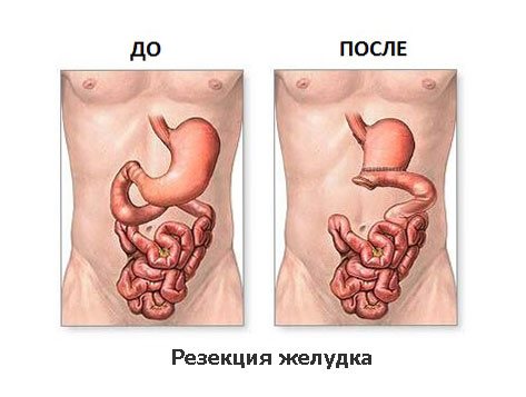
It can be of three types:
- resection – partial removal of the area of the main digestive organ;
- drainage - removal of liquid or gas from a seal;
- removal of the stomach. This method of surgical intervention is an extreme, undesirable measure. It is practiced only when there is a high probability of death.
From all of the above, a number of conclusions should be drawn:
- firstly, lumps in the stomach area are an extremely rare condition, in most cases having a congenital form;
- secondly, in most cases, compaction is treated with surgical methods;
- thirdly, with timely treatment, the risk of complications of the situation is unlikely.
If symptoms indicate the presence of lumps in the stomach, consult a doctor immediately. This will make it possible to avoid complications and cure the disease at an early stage.
People often try to independently examine themselves and make an accurate diagnosis. They often find a lump in their abdomen. If any formation appears, you must urgently consult a doctor and undergo a thorough examination, because this may indicate the development of serious problems.
Where can I get tested?
The first person to visit when diagnosing is a local doctor. He will conduct a superficial examination and question the patient. If necessary, the doctor will give a referral to a gastroenterologist. And only this specialist can prescribe medications and send for laboratory tests.
The second option is to go to a paid clinic or laboratory. There you can donate blood and feces for a fee to obtain a medical opinion. To prevent gastritis, it is imperative to do regular gastrointestinal checks.
Lump in the abdomen on the right side
If a lump is felt in the right side of the lower abdomen, then perhaps the reason lies in the formation of a formation in the area of the right kidney or ascending colon.
A lump on the right side of the lower abdomen signals the occurrence of certain diseases such as:
- appendicitis. The inflammatory process begins on the right side, but gradually moves to the navel and lower abdomen. Usually the disease is accompanied by acute pain, severe discomfort, fever and nausea. Urgent surgical intervention will help solve the problem;
- lipomas. In medicine it is also called benign wen. Occurs as a result of blockage of the sebaceous glands. On palpation, a soft tumor is felt. It can increase in size and roll under the skin. Removed by surgery;
- atheromas. This concept is usually understood as a cyst up to 3 cm in size, which is tightly attached to the skin. It is characterized by an inflammatory course, so it is hot and painful to the touch. Requires surgical removal;
- fibrosarcomas. A malignant formation, the diameter of which ranges from 2-15 mm. Resembles a smooth scar or bump on the skin. Characterized by mobility and pain. The middle part of the abdomen is tense, and tissue atrophy occurs over time. Treatment involves surgery;
- diverticulitis as a hernia with accumulation of intestinal contents as inflammation develops. Provoking factors are parasites, hereditary predisposition, poor nutrition, and decreased muscle tone. The disease is accompanied by upset stool, the passage of stool mixed with blood, an increase in temperature, and pain in the inflamed area. Treatment involves taking antibiotics. In complicated cases, surgical intervention is performed.
Lump in the stomach: treatment, causes, symptoms and types
/ From 23 to 27 years old
Back
: 02.03.2020
0
8
- 1 Alarm symptoms 1.1 Retroperitoneal
- 2 Seal under the skin in the form of balls
- 3 Why the appearance of such formations is dangerous
- 4 Lump on arm or leg
- 5 On the face
- 6 Diagnostics
- 7 Causes of lumps in the abdominal area
- 8 How to treat a wen (lipoma)
- 9 Seal on the joint
- 10 How to get rid of lipoma?
- Appendicitis. Inflammation of the appendix is manifested by acute pain on the right side, fever, and nausea. If this condition develops, a person must urgently contact a surgeon to perform an operation.
- Diverticulitis. The disease is accompanied by protrusion of part of the intestinal walls towards the abdominal cavity. This problem can be caused by helminthic infestations, unfavorable heredity or poor nutrition. The pathology is accompanied by indigestion, pain, and possibly increased body temperature.
- Aortic aneurysm located in the peritoneum. A similar problem arises against the background of hypertension, bad habits, and atherosclerosis.
Causes of lumps in the abdominal area
Lumps in the right side of the lower abdomen in women are most often caused by problems with the kidneys and large intestine. The causes of the appearance of formations on the left are called pathologies of the ureter and omentum. Intestinal pathologies are accompanied by lumps in the lower abdomen or in another part of it. Most often, lumps in the right or left side occur due to the following health problems:
Painful lumps can also occur with intestinal obstruction. This problem is accompanied by bloating, stool retention, vomiting, and severe pain.
Hydronephrosis is another common cause of lumps in the abdominal area. The disease is accompanied by accumulation of urine in the renal pelvis. It develops against the background of urolithiasis or when women have gynecological problems. In the presence of the disease, obstructed urine outflow, increased body temperature, and severe pain are observed.
Associated symptoms
It is difficult to say from the photo and external symptoms what could have contributed to the formation of the compaction.
First of all, after visiting a doctor, he collects information about accompanying symptoms, which may be the following:
- with an umbilical hernia: pain in the area of compaction, nausea, pain in the rectum, constipation;
- with diverticulitis (protrusion of the intestine, similar in appearance to a hernia): pain, fever, diarrhea alternately with constipation, the presence of blood in the stool;
- with a benign tumor: an increase in the size of the tumor, pain is often absent, as well as other unpleasant symptoms;
- with a malignant tumor: pain, erosion, crusts over the tumor on the epidermis, peeling;
- with atheroma cyst: pain, inflammation of the skin, rapid growth;
- when the bladder is bloated: discomfort when urinating;
- with an abdominal aortic aneurysm: aching pain radiating to the spinal region;
- with duodenitis: pain, dizziness, increased dryness of mucous membranes, general malaise, hypertension.
Signs of tumors
A lump under the skin in the form of a ball on the abdomen can be a tumor - benign or malignant. Most often, women are diagnosed with the following formations:
- Lipoma. A mobile bump on the skin is most often a benign tumor that is filled with fatty tissue.
- Atheroma. The formation looks like a ball that fits tightly to the skin. A dark dot may appear on the surface of the lump in the middle, which indicates a blockage of the sebaceous gland. Such a bulge on the body is accompanied by inflammation, and therefore leads to pain and may be hot to the touch.
- Fibrosarcoma. A malignant tumor with a diameter of 2-15 mm. It looks like a small painful bump on the skin. If left untreated, it becomes crusted or eroded over time.
If a small lump appears on your stomach, this may indicate the development of fibroids. A benign tumor consists of connective tissue. It can appear on the skin, tendons, mucous membranes or on the surface of internal organs.
Causes of lumps around the navel
Pathological seals can occur in the navel area. The main reasons for their development are:
- hernia;
- umbilical ring fistula;
- cyst;
- metastases formed in the presence of stomach cancer.
In the lower abdomen under the navel, the appearance of compactions may indicate the presence of inflammatory bowel pathologies and urolithiasis, rupture of the walls of the diverticulum. Tension and pain also appear with increased gas formation in the large intestine.
Palpation of the stomach
The stomach is palpated in the epigastric region with four half-bent fingers of the right hand folded together. They are installed 3-5 cm below the xiphoid process parallel to the position of the greater curvature of the stomach (Fig. 56, a). A superficial movement of the fingers upward to the xiphoid process first creates a skin fold.
Then, as the patient exhales, the fingertips are immersed deep and, upon reaching the spine, they slide from top to bottom. While the fingers are above the stomach, you can feel the rumbling. It is caused by the movement of fluid and gases in the stomach, caused by deep sliding palpation. Respiratory excursions contribute to better palpation of the stomach.
Therefore, the patient must be asked to calmly and deeply “breathe with his stomach.”
During the exhalation phase, the greater curvature of the stomach goes up, and the examiner’s fingers move down and slide off a small elevation in the form of a step formed by a duplicate of the greater curvature, which is felt at this moment as a soft elastic arc-shaped roller located on both sides of the spine.
To find the greater curvature, you can use the “double-handed” palpation method (Fig. 56, b).
For this purpose, the fingertips of the left hand are placed on the terminal phalanges of the right hand and deep sliding palpation is performed with them.
Normally, the greater curvature of the stomach is located 3-4 cm in men, 1-2 cm above the navel in women and is palpable in 50-60% of cases. When the stomach prolapses, it may lie below the navel
Rice. 56. Palpation of the stomach: a, b - greater curvature in the usual way and the “double hand” method; c — by palpation auscultation; d - percussion method; d - with the patient in an upright position.
The correctness of finding the greater curvature is checked by comparing palpation data with the results obtained using other methods of studying the lower border of the stomach.
When palpating the stomach, in addition to determining its location, you should pay attention to the consistency, surface and shape, as well as the presence of pain. In healthy people, the surface of the stomach is smooth.
Thickening of the greater curvature and pain during palpation are observed with gastritis and peptic ulcer disease. If a stomach tumor develops, its shape and consistency change, and the surface becomes lumpy. This is better revealed by palpation of the stomach in an upright position of the patient (Fig. 56, e).
Source: https://polic-5.ru/neotlozhka/proshchupyvaetsya-kishechnik-sprava.html
Lump in the abdomen on the left side

When the abdomen is divided into 4 parts, the renal hilum, ureter, omentum, and intestines are located in the left section.
A lump in the left side of the lower abdomen may indicate the development of:
- intestinal obstruction. There is persistent asymmetrical bloating, nausea, vomiting, increased passage of gases, retention of stool for more than 3 days;
- abdominal aortic aneurysm. The walls expand and bulge. It is more commonly diagnosed in men. The causes are hypertension, atherosclerosis, bad habits, and parasitic or bacterial infection. In addition to the lump, a dull pain is felt in the left side, which can radiate to the lumbar area. In the initial stages, drug therapy is carried out. In complicated cases, urgent surgical intervention will be required;
- hydronephrosis. Kidney disease that leads to accumulation of urine in the pelvis. Doctors identify several main causes of this condition in the form of urolithiasis, tumor formation, and prostate adenoma. The disease is accompanied by high blood pressure, increased temperature, colic in the lumbar region, and difficult outflow of urine with stinging and burning.
A lump on the left side in men appears when there is a urinary tract infection, prostatitis, urethritis or pyelonephritis.
If something is palpable on the right, left side near the navel or in the middle, then the possible cause is:
- umbilical or inguinal hernia;
- formation of a fistula in the umbilical ring;
- formation of cysts;
- swelling of the walls of the small intestine; metastases in gastric cancer.
Then the patient begins to complain of colic, nausea, vomiting, and increased temperature.
Palpation of the stomach: technique, norm and deviations. Anatomy of the stomach
A compacted formation located on the right side of the navel can be detected during a physical examination of a person. The development of compaction proceeds absolutely without discomfort, forcing the owner to think about health problems.
It is impossible to give a clear answer to why a lump appeared near the navel without diagnosis. There may be more than one reason here.
Umbilical cord hernia
An indicator of the appearance of a hernia is a palpable dense area in the abdominal area, which does not disappear when pressure is applied to it. Pressing is accompanied by severe pain, and the hernia becomes even more noticeable.
The following symptoms are also observed:
- bloating;
- bowel dysfunction;
- nausea and vomiting;
- increased heart rate.
There are two types of umbilical hernia - strangulated and reducible.
If any of them appear, you should contact a specialist. The first type is eliminated through surgery, and the second type is eliminated by manual reduction of the seal that appears near the navel.
Dissection of lymph channels
Dissection of the lymph channels during the intervention can also cause a seal to appear above the suture. When the damaged lymphatic channel does not heal, fluid moves and penetrates into the free space in the abdominal cavity filled with lymph.
This phenomenon may occur after laparoscopy if the operation was performed poorly on the uterine appendages. In any case, the dissected tissue in the abdominal cavity is fastened and stitched with threads.
If the surgeon is inexperienced and negligent, and the quality of the operation is poor, complications arise, the development of bacteria, and infection of sutures and wounds.
At best, scar tissue will appear under the skin, and at worst, cells will begin to actively multiply in this area, and a keloid scar will form due to infection and the development of inflammation.
All women need to carefully monitor the sutures after surgery: take preventive measures and follow medical instructions, regularly treat the sutures with antiseptic solutions (brilliant, Fukortsin), and stick a plaster on the surface of the suture.
Alarming symptoms should be the reason for an early visit to the gynecologist: colic and severe pain in the incision area, ichor oozing from the wound, serous putrid odor, increased local temperature, swelling and redness in the stitching area, unpleasant bleeding from the vagina with pain, burning and itching . An examination is required, mainly in case of suppuration - cleaning of the sutures.
Lump in the upper abdomen
At the top left of the abdominal cavity are the spleen, diaphragm, stomach, pancreas and part of the intestinal loops. Some diseases can lead to enlargement of the spleen and rupture. The cause may be injury or impact. Then the person complains of severe pain and the appearance of a bluish lump near the navel.
There are other reasons why a tumor appears, such as:
- peritonitis;
- perforation of the ulcer;
- irritation of the mucous membrane of the stomach and duodenum;
- tearing or stretching of the tissues of the abdominal aorta;
- volvulus of the sigmoid colon;
- tumors of the intestinal canal;
- Crohn's disease.
Only an experienced doctor can make an accurate diagnosis and identify the cause of the formation of the seal.
Can the intestines be felt?
During palpation, the examining fingers are carefully immersed and pressed against the organ being examined to the posterior abdominal wall;
Contours, density and possible formations and deviations are determined by sliding movements. As a rule, when palpated, the sigmoid colon gives the impression of a smooth, dense, mobile, non-rumbling and painless cylinder the thickness of a finger. Its thickness depends on the condition of the walls, filling with gases and fecal matter.
With inflammatory infiltration, its walls thicken; when overflowing with solid fecal matter, the sigmoid colon becomes clearly shaped, and deep ulcerative processes make it lumpy and uneven. With an acute inflammatory process in the sigmoid colon, the latter acquires a denser consistency and becomes painful.
The density of the sigmoid colon, filled with gases and liquid contents, is reduced; when inflammatory adhesions form around it, normal mobility is lost. During spasm, the intestine can be felt in the form of a cord or cord.
Rumbling in the sigmoid colon occurs when liquid contents enter from the upper sections or when feces are retained for a long time; the latter entails irritation of the walls with the release of a significant amount of mucus (false diarrhea).
The cecum is normally palpable in the form of a smooth two-finger-wide, slightly rumbling, painless and moderately mobile (2-3 cm) cylinder. Its mobility can be pathologically increased (mobile cecum - coecum mobile).
The consistency thickens with coprostasis, distension by gases, acute and chronic inflammation, but the walls remain smooth and even. In the presence of a tuberous cecum, one should think about ulcers of tuberculous, syphilitic, dysenteric origin, or a tumor, penetrating deeply into the wall.
The volume and shape of the cecum depend on the quantity and quality of its contents. With thick contents and a normal amount of gases, the intestines do not rumble; with liquid contents in combination with a significant amount of gases, a loud rumbling occurs, most often with enteritis, typhlitis.
The presence of pain upon palpation of the cecum always indicates its pathological condition.
After palpation of the cecum and very rarely the appendix, they move on to palpating the less accessible parts of the large intestine - the ascending, transverse colon and descending colon. The transverse colon can be palpated only when it is chronically inflamed.
Consistency, volume and shape depend on the tone and degree of tension of its muscles, as well as the properties of the contents. Any inflammatory process, especially ulcerative, in the presence of inflammatory infiltration causes serious changes in the transverse colon.
It changes its shape and consistency, its walls thicken, under the influence of the ulcerative process the muscles contract more strongly, and its configuration changes.
With chronic colitis and pericolitis, the intestine becomes dense, contracted, painful on palpation, and sometimes lumpy (at the site of ulcers). With pericolitis, it loses both respiratory, active and passive mobility due to the adhesions that form.
By palpating the abdomen, you can feel an intestinal tumor, which is often confused with a tumor of neighboring organs. Tumors of the transverse colon and cecum are characterized by known mobility. Tumors of the transverse colon and its flexures move during breathing, while tumors located below the navel are usually motionless.
With enterocolitis, palpation of the abdomen causes rumbling and splashing noise in the navel area.
The small intestines can be felt mainly near the navel. With enteritis, painless diarrhea is noted, and upon palpation of the small and large intestines - rumbling. With colitis, mushy, mucous stools, abdominal pain are observed, and upon palpation, a painful, compacted, dilated and slightly rumbling colon.
Palpation of the abdomen is supplemented by digital examination of the rectum, sigmoidoscopy and x-ray examinations. For all intestinal diseases, a digital examination of the rectum should be performed so as not to miss rectal cancer and syphilitic structures.
Digital examination in combination with sigmoidoscopy allows us to determine the presence of inflammatory processes, cracks, fistulas, tumors, and hemorrhoids. In addition, an impression is made of the tone of the sphincter, the width and filling of the rectal ampulla.
In some cases, palpation of adjacent organs is very useful - the pelvic floor, the pouch of Douglas, the neck and bottom of the bladder, in men - the prostate gland and seminal vesicles, in women - the uterus and its appendages.
A digital examination can detect a tumor of the rectum and sigmoid colon; in women, a tumor of the uterus and an ovarian cyst, which are directly adjacent to the rectum, compressing or pushing it to the side.
Digital examination sometimes makes it possible to determine the nature of constipation. It is known that under normal conditions the ampulla of the rectum is empty, but with chronic constipation due to impaired innervation of the muscular system, it can be overcrowded and expanded.
ABDOMINAL ORGANS: DEEP PALPATION OF THE ABDOMEN
After superficial palpation of the abdomen, the abdominal organs accessible through deep palpation are examined, determining their position, size, shape, consistency, surface condition, and the presence of pain. In this case, additional pathological formations may also be detected, in particular tumors and cysts.
The research conditions are the same as for superficial palpation of the abdomen. To reduce tension in the abdominal muscles, you need to ask the patient to slightly bend his knees so that the soles of his feet are completely on the bed. In some cases, palpation is additionally carried out with the patient in an upright position.
To clarify the boundaries of individual organs, along with the palpation method, percussion and auscultation are used. In addition, in order to identify pain in the projection of organs that lie deep in the abdominal cavity and are inaccessible to palpation, penetrating palpation is used.
In patients with ascites, the method of ballot palpation is used to examine the abdominal organs.
One of the most important conditions for deep palpation of the abdominal organs is knowledge of their projection on the anterior abdominal wall:
- left hypochondrium
: cardia of the stomach, tail of the pancreas, spleen, left flexure of the colon, upper pole of the left kidney; - epigastric region
: stomach, duodenum, body of the pancreas, left lobe of the liver; - right hypochondrium
: right lobe of the liver, gall bladder, right flexure of the colon, upper pole of the right kidney; - left and right lateral areas (flanks of the abdomen)
: respectively, the descending and ascending sections of the colon, the lower poles of the left and right kidneys, part of the loops of the small intestine; - umbilical region
: loops of the small intestine, transverse colon, lower horizontal part of the duodenum, greater curvature of the stomach, head of the pancreas, renal hilum, ureters; - left iliac region
: sigmoid colon, left ureter; - suprapubic region
: loops of the small intestine, bladder and uterus when they are enlarged; - right iliac region
: cecum, terminal ileum, appendix, right ureter.
Source: https://limto.ru/mozhet-li-proshhupyvatsja-kishechnik/
Lumps in the lower abdomen
A pathological formation can appear in any part of the abdomen. If a lump is felt in the lower abdomen, then the cause is:
- pinched hernia;
- protrusion of the sigmoid colon;
- rupture of the walls of the diverticulum;
- inflammatory process in the pelvic organs;
- intestinal obstruction in the lower part of the organ;
- increased gas formation;
- tumors in the male genital organs.
Irritable bowel syndrome, an inflammatory process in the intestinal canal, or compression of the nerves can lead to the appearance of a lump in the lower abdomen. A lump often occurs as a stone moves down the ureter.
Pain on palpation
Palpation of the digestive organs is of great diagnostic value. The formation of a symptom is influenced by a number of factors and nuances (infection, structure of the reproductive system).
Features of pain on palpation
The process of palpation of the stomach and intestines is important in terms of diagnostic examination of the human body.
Monitoring of the digestive organs is carried out as follows: at the first stage, a qualified specialist carefully palpates the sigmoid colon - this is the most common landmark and the most accessible organ for palpation.
Next, the doctor proceeds to study the condition of the cecum and transverse colon. The ascending and descending sections of the suction organ are quite problematic to palpate.
In practice, during palpation, fingers must be carefully immersed on the surface of the body area and gently pressed onto the organ being examined (toward the posterior abdominal wall). Using sliding movements, you can clearly determine the contours, density, and the presence of various neoplasms and abnormalities.
When you touch (feel) the sigmoid colon, you get the impression that there is a smooth, dense and mobile cylinder in the human body. The size of such a “geometric figure” does not exceed the thickness of a person’s thumb.
The formation parameters are directly related to the condition of the walls, which are densely filled with gases and decay products (feces/feces).
During the inflammatory process of the infiltrating walls, a significant thickening of the membrane occurs. Ulcerative manifestations form a lumpy and uneven surface of the suction organ.
Acute inflammation of the sigmoid colon is accompanied by the formation of a dense consistency of painful manifestation. Due to the dense overflow of gases and liquid contents, motor inhibition occurs. The spasm is felt in the form of a cord and cord.
The patient experiences systematic rumbling + false urge to defecate (false diarrhea).
In normal condition, the cecum is easily palpable. A specialist can detect a cylinder up to 3 cm that is moderately active in movements. Its mobility in a pathological disorder is significantly increased.
The internal consistency becomes significantly thicker during coprostasis and chronic inflammation. The volume and shape of the cecum directly correlates with the contents. In a normal functional state, the intestines do not growl.
The patient should remember that the presence of pain upon palpation in the area of the cecum indicates the development of a pathological process. The digestive organ requires systematic and comprehensive treatment.
In practice, after examining the cecum (+ appendix), it is possible to examine the less accessible parts of the large intestine. Palpation is carried out from the ascending to the transverse colon and descending colon.
The transverse colon part of the suction organ can be qualitatively palpated only in the case of chronic inflammation. Tone, consistency, volume, shape depend on the tone and degree of muscle tension.
For example, an inflammatory process of the ulcerative type forms serious preconditions for the transformation of the transverse colon. At the same time, the muscles of the organ thicken significantly, and its configuration changes.
Today, chronic colitis and percolitis are quite common. With these ailments, the wall of the suction organ begins to painfully contract. Due to the lumpy surface, palpation is accompanied by sharp pain. For example, with pericolitis, respiratory and active mobility is lost.
Palpation of the abdomen allows you to feel an intestinal tumor, which is often confused with the pathology of various organs. Oncology of the cecum and transverse colon is distinguished by already known mobility. The pain is activated during the act of breathing (tumors below the navel are motionless). Palpation of the abdomen during enterocolitis is accompanied by rumbling in the navel area.
The disease has specific signs and symptoms: painful diarrhea (mushy, mucous stools, abdominal pain, hard colon). Palpation of the abdomen is carried out in combination with a digital examination of the rectum (sigmoidoscopy + radiography). These actions make it possible to predict the formation of rectal cancer and the formation of various syphilitic structures.
It will also be possible to clearly determine the presence of inflammatory processes, cracks, fistulas, hemorrhoids and all kinds of tumors. The specialist can get a clear vision of the sphincter tone and the level of filling of the colon ampulla. In some cases, it is rational to palpate adjacent organs (bottom of the bladder, prostate gland, uterus with appendages).
This will reveal an ovarian cyst, a tumor of the genital organs, the degree of constipation, etc.
Mechanism of the procedure
Palpation is the last stage of a full and objective examination of the abdominal area. The patient will need to cough vigorously before the procedure. In practice, a person with developed peritonitis manages to do this only superficially (holding his stomach with his hands).
It is allowed to make a small impact on the couch on which the patient is located in a supine position. The vibration impulse will provoke the manifestation of pain in the gastrointestinal tract. Thus, it is quite easy to establish the diagnosis of peritonitis without touching the hand.
To identify symptoms of peritoneal irritation, it is allowed to gently shake the patient after grasping the crests of the ileum (or jumping on one leg).
The palpation procedure begins with the patient being asked to clearly indicate the area where the first pain formed (the primary localization of the disease).
The specialist needs to closely monitor the actions of the patient himself. This is how you can identify the causes of peritoneal irritation.
Diffuse visceral pain in the abdomen is easily determined using circular movements of the palm. Your hands should be warm.
The procedure begins as far as possible from the main source of pain. This helps to avoid unplanned pain at the very beginning of the study. Children, and sometimes adult patients, sometimes prevent a quality examination due to pain.
First of all, the doctor must perform gentle and careful palpation (superficial). An experienced specialist moves gently, methodically and consistently. The fingers make a minimum number of movements.
It is strictly forbidden to palpate the abdomen randomly! The pressure on the body surface should not be high. Otherwise, protective tension in the abdominal muscles will occur.
Touching the sore spot must be done until the patient says that it really hurts.
A qualified specialist can always determine the degree of tension in the muscles of the anterior abdominal wall. The physician must distinguish between voluntary and involuntary muscle tension. To clearly determine this factor, during palpation a person takes a deep breath and exhales. If muscle activity persists, this indicates the development of peritonitis.
It is rational to carry out deeper palpation if peritonitis was not detected during a superficial examination. This makes it possible to detect various tumor formations, hepatosplenomegaly, and aortic aneurysm. It is very important for a physician to remember the optimal sizes for normal structures so as not to confuse them with malignant ones. Pain on palpation of the abdomen and intestines has two types:
- immediate local pain – the patient experiences sharp pain at the test site;
- indirect (referred pain) – pain sensations are formed in a different place when palpated. For example, during acute appendicitis, pain accumulates at McBurney's point on the left side of the iliac fossa. This symptom is called “Roving” and is a reliable sign of peritoneal irritation.
It is easy to carry out comparative palpation of a patient with tense abdominal muscles. For this, the patient, who is in a supine position, is asked to gently lift his head above the pillow.
The classic symptom of parietal peritoneal irritation is not difficult to identify.
To do this, at the time of the examination, the doctor must sharply remove his hand from the surface of the body and observe the patient’s reaction. In most cases, patients experience a significant increase in pain.
This classic examination technique is quite crude; some scientists classify it as a barbaric method of study.
With the development of various pathologies in the digestive organs (for example, acute appendicitis), hyperesthesia of the skin in the abdominal area is observed.
It is for this reason that if you pinch or lightly prick a patient, a painful reaction of the body will immediately occur.
This is a fairly common clinical symptom, but its establishment is not enough to firmly diagnose acute appendicitis and other diseases of the abdominal organs.
An integral part of the palpation examination is gentle tapping on the lumbar region (+ sides of the abdomen) to determine the degree of pain in these areas. Quite often, pyelonephritis and urolithiasis correlate with severe pain in the abdomen (costovertebral region).
In doubtful clinical situations, examination alone is not enough. An accurate assessment of the dynamics of the disease is established by repeated palpation of the abdomen by the same doctor.
Causes of pain in women
Today, medicine identifies two types of fundamental causes that affect pain when palpated. Organic factors include:
- inflammatory processes in the genitourinary system (cyst, endometritis, fibroids);
- using the IUD as a contraceptive;
- formation of various pathological formations;
- the presence of inflammation in the gall bladder (including appendicitis, pyelonephritis);
- sharp pain during pregnancy (placental abruption, miscarriage).
The functional reasons are as follows:
- systematic disruptions in cycles during menstruation;
- discharge of uterine bleeding;
- ovulation + uterine inflection.
Inflammatory processes are the main cause of pain during palpation of the stomach and intestines. The disease begins with classic acute manifestations and is supplemented by various signs of intoxication of the body, namely:
- Endometritis is accompanied by aching pain in the abdominal area. Their manifestation can be determined by light palpation. The patient experiences heaviness in the area of the appendages + hardening of the uterus;
- Endometriosis is a pathological disorder that affects the uterus and appendages. Severe pain is observed when palpating the middle of the abdomen;
- Ovarian apoplexy correlates with ovulation. In this case, some of the blood penetrates into the abdominal cavity due to strong physical exertion;
- Uterine fibroids. The pain syndrome is localized in the lower abdomen (compression of neighboring organs);
- Appendicitis requires surgical medical intervention. Pain on palpation in the area where the appendix is located;
- Cholecystitis is an inflammatory process of the gallbladder. The pain radiates clearly to the lumbar region and back;
- Cystitis is a lesion of the bladder. Pain is observed both during palpation and during urination.