What you should remember for the correct X-ray of the stomach with barium
Very often, doctors cannot make an accurate diagnosis, since many diseases of the gastrointestinal tract have similar symptoms. Therefore, the patient is prescribed an x-ray of the stomach and esophagus. It can be used to identify abnormalities in the structure of organ walls and the presence of possible formations. The main indications for conducting such a study are:
- severe pain;
- nausea and vomiting;
- difficulty swallowing food.
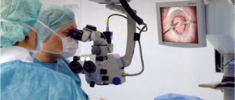
- impossibility or presence of contraindications for FGDS (fibrogastroduodenoscopy)
Before carrying out the procedure, it is very important to prepare properly; only in this case can you obtain reliable results. “Preparing” the patient before an x-ray of the esophagus includes a strict diet and limitation of physical activity. Let's look at this in more detail.
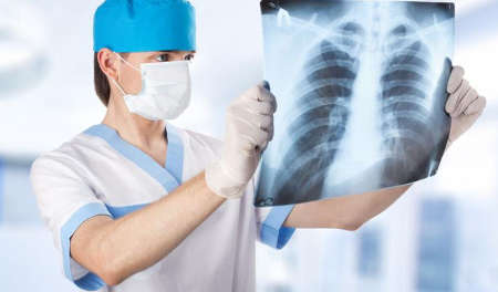
What is radiography of the esophagus: indications and methods
Timely detection of the disease greatly increases the chances of a full recovery.
This article will discuss in detail the diagnostic procedure called radiography of the esophagus and stomach, indications for its implementation, preparation rules, results and much more. Radiography is a special method for studying the internal organs of a person, thanks to which doctors can obtain the desired image on a photograph or x-ray film.
Radiography has two main types:
- Diagnostic method with contrast. This means that during the examination the patient must take a special contrast agent, thanks to which his internal organs will be much more clearly visible in the finished image.
- The double contrast method involves the procedure with barium. Most often, this method is used to assess the elasticity of the walls in the patient’s stomach.
X-ray of the esophagus and stomach is prescribed if the following diseases and pathologies are suspected:
- Stomach ulcer.
- Tumor of the esophagus or stomach.
- Gastritis in the stomach.
- Enteritis.
- Colitis.
- Duodenal ulcer.
- Pathologies of stomach development.
- Diverticula.
- Monitoring the condition of the esophagus or stomach after surgical treatment.
- Stenosis.
- Scarring.
- Pathologies of the esophagus.
- Suspicion of a foreign object entering the esophagus.
In addition, direct indications for X-rays of the esophagus and stomach are the following symptoms:
- sharp pain when swallowing;
- pain in the esophagus or stomach;
- burning sensation when swallowing;
- belching;
- pain in the navel area;
- indigestion;
- nausea.
This study is contraindicated in the following cases:
- pregnancy, especially in the first stages of its course (this study cannot be carried out so as not to harm the child and its development);
- serious condition of the patient;
- prolonged stomach bleeding;
- individual intolerance by the patient to the contrast agent;
- allergy to iodine.
Most often, diseases in these organs appear for the following reasons:
- Eating fatty, too spicy or fried foods.
- Binge eating.
- Alcohol abuse.
- Smoking.
- Eating food with harmful chemical additives.
- Burn or chemical damage to the mucous membrane of the stomach and esophagus.
- A foreign object enters the esophagus, causing damage to its mucous membrane.
Preparation for radiography includes the following measures:
- Twelve hours before the examination, the patient should not eat anything so that the doctor can better see the stomach cavity in the image. If the diagnosis is carried out on an infant, then it is also advisable not to feed him until the contrast liquid is administered.
- The patient needs to be explained in detail how the x-ray will be performed and what needs to be done for this.
- The subject is asked to drink barium contrast, which may taste a little strange. At the same time, you should know that first he must drink a liquid that is denser in consistency, and then diluted in a volume of 350 ml.
- The patient is warned that x-rays should be taken in only one position of the body, so the person should not move.
- If esophageal reflux is suspected, the person should refrain from taking medications the day before the procedure.
- Immediately before starting the procedure, the subject must remove all metal chains, dentures, hairpins, etc.
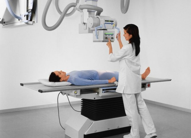
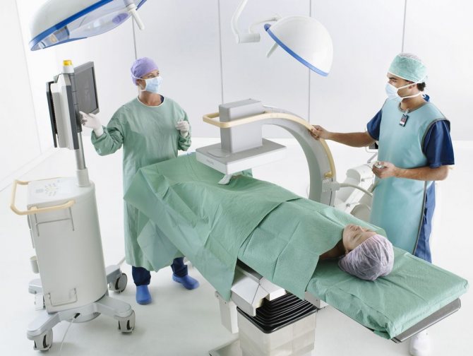
Radiography is performed in the following sequence:
- The patient stands straight behind the X-ray screen.
- The doctor takes an image of his stomach and esophagus.
- Next, the patient drinks several sips of barium contrast. Sometimes the progress of this swallow is also recorded to better see how the liquid will move down the esophagus.
- After this, side views are taken.
Typically, young children drink barium contrast without resistance, but if the child does not want to drink this substance, then it is injected into his stomach using a small medical tube.
When examining infants, it is customary to use special moving platforms. This will allow the radiologist to see the baby's internal organs more clearly.
In adolescents, double contrast studies are often performed.
- Next, the patient lies down on the table and pictures are taken in a supine position (this helps to identify a hiatal hernia, as well as esophageal reflux).
- Typically, the total duration of such an examination is 20 minutes.
After examining the patient’s completed images, the doctor will draw up a detailed report, which is then sent to the attending physician. Based on the information received, the doctor will select the necessary treatment for each patient individually (depending on the diagnosis, age and condition of the patient).
It is important to know that it is almost impossible for an ordinary person without special medical education to accurately interpret X-ray images, so it is better to entrust this to an experienced specialist.
Additional pathologies that may be detected after radiography:
- Changes in the lumen of the stomach or esophagus. This concept means a narrowing or expansion of the lumen of organs with growing oncological pathology.
- Dilation of the lumen can also occur with diverticula.
- Deformation of the organ under study.
- Violation of the structure or integrity of an organ.
- Filling of one or more areas of the organ with a contrast agent is characteristic of a stomach ulcer.
This examination has the following advantages:
- Helps to diagnose the disease in a timely manner, thereby increasing the patient’s chance of proper treatment and a speedy recovery.
- X-ray is a relatively safe and painless procedure.
- The study allows you to view the condition of the patient’s internal organs as accurately as possible.
- Does not cause any allergic reactions.
- Does not leave active harmful radiation after the procedure.
- Does not cause any side effects.
Despite a number of advantages, radiography also has the following disadvantages:
- If radiography is performed frequently, it increases the risk of developing cancer pathologies in the body.
- The required radiation dose is different for each patient.
- Very rarely, patients are allergic to flavoring additives that are mixed into the barium liquid.
- There is a risk of barium retention in the intestines, which can cause short-term intestinal obstruction.
- X-rays should not be performed during pregnancy.
- Sometimes after this procedure, patients complain that the consistency of the barium contrast is too thick, making it difficult to drink. In addition, liquid barium has a strong chalky taste, making it very difficult to drink without fruit flavors.
- Some discomfort for the people being examined is caused by the need to change their body position during the procedure.
- You may experience nausea and bloating after taking barium.
- When using special liquid soda, a person may experience nausea and bloating.
- Two days after the procedure, the stool may become grayish in color due to the presence of barium in the body.
- Sometimes after taking this contrast, people suffer from constipation and indigestion, so it is recommended to drink plenty of fluids in the first days after the x-ray.
- Give up bad habits (drinking alcohol, smoking).
- Eat properly.
- Eat small portions 4-5 times a day.
- Drink plenty of fluids.
- Do not eat too hot food.
- Chew every bite of food thoroughly.
- Avoid stress.
- Keep a food diary in which you write down everything you eat. This will control the menu and protect against the desire to eat “something tasty, but at the same time harmful.”
Today, radiography is considered a very effective procedure for diagnosing a wide variety of pathologies of the stomach and esophagus. Immediately after the initial examination of the patient, the doctor prescribes it in order to see the complete condition of the internal organs.
In addition, experts recommend X-rays for preventive purposes at least once a year. This will help to timely identify the disease at the initial level of its development, which will greatly simplify treatment.
In gastroenterology, two types of contrast are used to diagnose diseases of the esophageal tube: conventional and double contrast methods. In addition, there are two subtypes of the procedure: radiography of the esophagus and stomach, as well as fluoroscopy of the same organs. Despite the similarity of the methods, they still have fundamental differences.

It is common practice to use barium for X-rays of the stomach and esophageal tube, since hollow organs are not visualized well enough. This is a contrast agent consisting of a suspension of barium sulfate salts in drinking water. Externally, the solution looks like a milky-white liquid with a neutral chalky taste. When examining the stomach and esophagus with barium, specialists are able to examine in detail the internal outlines of the organs.
Important! When examining the stomach with barium, the drug is not absorbed, so the method is considered absolutely safe in terms of systemic effects on the body.
X-rays with contrast, in addition to better visualization of the internal contours and structure of the mucous membrane, make it possible to evaluate the peristalsis of organs and other aspects of their functioning. To conduct an informative study of the stomach, contrast is drunk immediately before taking a series of images.
Although barium is considered a safe compound by radiologists, patients may experience allergic reactions to barium sulfates. If this happens once, in subsequent studies a different contrast is used, most often iodine compounds.
Principles of preparation
Preparation for fluoroscopy includes:
- avoiding taking any medications;
- alcohol and tobacco products are prohibited;
- the last meal should be no later than 7-8 hours before the procedure;
- in the morning before the analysis, it is forbidden to eat or drink;
- It is imperative to prepare the intestines with a cleansing enema (nowadays special microenemas or mild laxatives are very often used).
The patient should begin preparing a week before the procedure. It is necessary to exclude all fried, spicy and fatty foods, carbonated drinks and foods that contribute to increased gas formation. Limit intake of dairy products and heavy foods (this applies to meat). During the day you need to eat only steamed or boiled dishes with a minimum of salt. If the patient was unable to strictly adhere to all conditions, then a repeat test is scheduled after 7 days.
Preparing for the examination
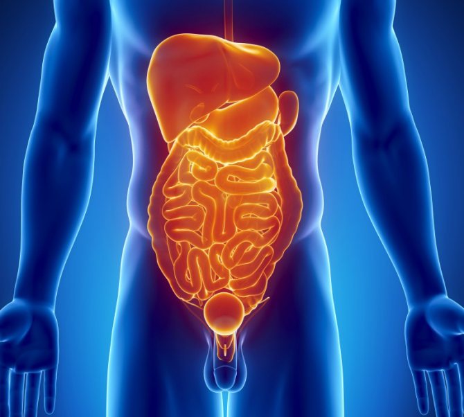
The stomach is a hollow organ. To achieve the best quality images, it needs to be filled with a special liquid that will not transmit ionizing radiation during the study. An aqueous suspension of barium sulfate is used as a dye. Double contrast is also used, when air is introduced into the stomach in addition to barium under pressure.
This technique makes it possible to diagnose many pathologies of the stomach and duodenum, and also allows you to identify a hiatal hernia. Preparation for x-rays of the intestines and stomach involves dividing patients into two conditional categories:
Without disruption of digestive function
Such patients only need to avoid eating 8 hours before the start of the study. The procedure is carried out strictly on an empty stomach. Also, on the eve of the procedure, you should avoid drinking alcohol and quit smoking. Do not take medications or use chewing gum. The latter stimulates the secretion of saliva and hydrochloric acid, which can prevent dyes from enveloping the mucous membrane of the organ being examined.
With disorders of the digestive tract
Preparation for fluoroscopy in patients with such problems is more serious. 2-3 days before the test, sick people must follow a special diet. It includes avoiding foods that can cause increased gas formation (carbonated drinks, whole milk, legumes, fresh baked goods, etc.).
If flatulence still makes itself felt, you can take medications such as Espumisan and activated carbon. If constipation is present, patients are given an enema the day or several hours before the test. In some cases, gastric lavage with a probe is required. This procedure is performed for stenosis of the pyloric region of this organ.
Technique
2 hours before the start of the study, the intestines are cleansed, since an X-ray examination of the stomach is carried out with an examination of the intestines the next day. Then the patient is given a special enveloping solution to drink, which includes barium sulfate. It plays the role of a contrast agent. It is recommended to remove all jewelry that may distort the picture.
The patient is examined in three positions: standing, lying down and the Trendelenburg position (pelvic elevation by 450). The doctor takes a series of pictures in different projections. In order for the contrast solution to be evenly distributed, the doctor performs a small massage in the abdominal area.
Sometimes a procedure may be prescribed using a double contrast solution, which includes barium, almagel, sorbitol, sodium citrate, carboxymethylcellulose and ethanol. The patient drinks it through a special straw.
It is important to note that during this procedure the patient is heavily exposed to radiation, so it is not recommended to undergo it more than once a year.
Symptoms that indicate diseases of the stomach and gastrointestinal tract
Of course, a specialist can prescribe an x-ray of the stomach with barium, but the patient himself must clearly understand that at the first symptoms it is necessary to immediately consult a doctor in order to accurately determine what stage the disease is at and provide timely treatment. The following symptoms may be prerequisites for a barium x-ray:
- A person loses weight very quickly.
- There is often a burning sensation in the stomach area.
- Belching occurs for no reason.
- Problems with the swallowing reflex appear.
- There is severe pain in the navel area.
- When defecating, you can see blood clots.
At the slightest manifestation of symptoms, it is recommended to immediately consult a specialist.
Contraindications for carrying out
This procedure is not recommended in the following cases:
- individual intolerance to a component of the contrast solution;
- severe and prolonged internal bleeding;
- pregnancy in the early stages (prescribed in the 2nd or 3rd trimester only for special indications).
In some cases, doctors may, as an exception, perform fluoroscopy if the risk of negative consequences is low.
Features of the examination
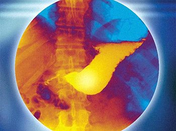
An X-ray of the stomach with barium is an extremely informative procedure that allows you to assess how normal the gastric mucosa is.
Depending on the results of the study, the doctor will be able to determine the presence of a variety of pathologies, and both major damage and the most minor deviations will be visible.
Another name for a barium X-ray of the stomach is double contrast.
Photo:
Before starting the procedure, the patient will have to drink a special mixture that will color the walls of the stomach and help the doctor identify damaged areas.
The contrast mixture consists of 700 grams of sodium sulfate dissolved in hot water. Almagel is also added to the solution - it is necessary to make the solution more viscous.
Other agents are also added to the composition, for example, sorbitol, antifoam, sodium citrate and other compounds necessary to create an optimal environment in the stomach for research.
The solution is prepared immediately before the start of the procedure. The patient needs to drink about a third of a glass. This element is inserted using a special tube with a hole.
However, in case of intestinal perforation or suspicion of this pathology, barium is replaced with gastrografin without fail for safety reasons.
After administering the solution, the doctor can begin radiography. During the procedure, six pictures are taken, and the patient will need to change position several times - this is necessary so that the doctor can examine the organ from all sides.
Pictures are taken with the patient lying down, standing, and with the pelvis raised at an angle of 35 degrees.
Photo:
During an x-ray, the device makes a left lateral and standing projection, and also examines the antrum and lateral wall in an oblique projection.
In addition to the stomach, this procedure also allows you to take pictures of the duodenal bulb.
READ Features of intestinal x-ray
Despite the obvious benefits of the study, such an x-ray has negative consequences, since the patient receives a fairly noticeable dose of radiation. For this reason, conducting research more than once a year is prohibited.
Possible consequences
The patient may experience negative consequences after fluoroscopy:
- constipation due to increased viscosity of stool by the contrast agent component;
- discomfort during hunger;
- change in stool color during the day;
- increased gas formation, nausea, discomfort in the abdomen.
If any of these signs are present, it is recommended to immediately consult a doctor for advice. In order to reduce the risk of developing consequences, the patient must adhere to strict diet for a week after the procedure. In this case, exclude all foods that can bind stool, increase the amount of gases, and drink plenty of fluids. It is recommended to enrich your diet with dairy products, fresh vegetables and fruits. The consumption of alcohol and tobacco products is strictly prohibited. You need to walk in the fresh air more often and do light exercise.
Thus, a roscopic or graphic examination of the stomach followed by examination of the intestines allows one to assess the condition of the mucous membranes of the digestive system. Thanks to this technique, gastroenterologists can accurately diagnose the patient. It is very important to properly prepare for such a study; this will improve the image in the image and obtain reliable results. Negative consequences are rare. To reduce the risk of their development, you must follow all medical recommendations.
Fluoroscopy analysis of the stomach
The results of fluoroscopy of the stomach are interpreted by a radiologist and gastroenterologist, and sometimes by a surgeon. With fluoroscopy, you can obtain data on the speed of movement of barium suspension through the esophagus and stomach, and the time of its entry into the duodenum and colon. Based on this sign, one can judge the presence of motor disorders and difficulties in evacuating a food bolus from the esophagus or stomach. Based on the relief of the mucous membrane and the uniformity of barium distribution, the presence or absence of its changes is determined.
Symptoms determined by fluoroscopy of the stomach
- Changes in the position of the esophagus and stomach (with congenital anomalies of the structure of the digestive organs, prolapse of the stomach, previous operations, tumors of the mediastinal organs, myocardial hypertrophy).
- Narrowing of the lumen of the esophagus or stomach (tumors, strictures, cicatricial narrowings due to ulcers, chemical burns, increased tone of the muscle wall).
- Expansion of the lumen (with achalasia cardia - expansion of the esophagus, decreased wall tone with Hirschsprung's disease and functional disorders).
- Violation of the integrity of the wall (perforation of the walls of the esophagus or stomach due to ulcers).
- Symptom of “niche on the contour” (ulcers).
- Filling defect (polyps, papillomas, foreign bodies, malignant tumors).
- Reducing the folding of the mucous membrane (with atrophic gastritis).
- Radiation-like convergence of mucosal folds (together with the niche symptom indicates a peptic ulcer).
In what cases are x-rays of the esophagus and stomach taken?
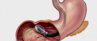
X-ray of the esophagus and stomach is often prescribed for diseases of the gastrointestinal tract, as an additional or primary, and often the only possible method of examination (when it is impossible to insert an endoscope due to organic obstacles or the patient’s panicky fear of the examination).
Considering that the procedure involves x-ray radiation, it can only be prescribed by a doctor.
Existing methods of X-ray examination (general X-ray, X-ray with simple and double barium contrast, CT examination, including multispiral CT, fluoroscopy), various positions of the patient (Trendelenburg) make it possible to identify almost any pathology of the esophagus and stomach. To examine the upper esophagus and larynx, G. M. Zemtsov’s technique is used.
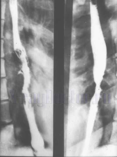
X-rays of the esophagus and stomach are performed using barium contrast, which fills not only the esophagus and stomach, but also the intestines. Thus, with one examination you can see the condition of the entire digestive tract. Barium resembles a mushy mass, the consistency of sour cream, white in color, and tastes like chalk.
Types of fluoroscopy of the esophagus
X-ray examination of the esophagus and stomach is an optimal procedure that is easily tolerated by the patient. It is used even in seriously ill patients when endoscopy is difficult. With fluoroscopy, all segments of the digestive tract, parts of the stomach are clearly visible; you can examine the duodenum, small and large intestines.
The main types of simple x-ray diagnostics are:
- survey fluoroscopy;
- radiography with contrast;
- double contrast radiography;
- fluoroscopy.
Plain radiography of the esophagus and stomach
Plain radiography, as a separate diagnostic method, is now practically not used due to its low information content. The fact is that the density of the upper gastrointestinal tract is almost the same as that of the surrounding tissues (heart, liver, lungs). Therefore, in the picture, the esophagus and stomach look like a poorly defined spot that is difficult to describe.
Sometimes the attending physician asks the radiologist to look at the condition of the mediastinum and the relative position of the organs, then a survey radiograph is irreplaceable.
In addition, survey fluoroscopy is the mandatory first stage of X-ray diagnostics of the gastrointestinal tract before the patient receives contrast.
X-ray of the esophagus and stomach with barium
In order to better examine the esophagus and stomach during radiography and identify their pathology, various contrast agents are used. There are a large number of contrast agents available, the most commonly used is barium sulfate. If one contrast agent is intolerant, it can be replaced with another, more suitable one. If fistulas, failure of the sutures of the operated esophagus, or other destruction of the esophageal wall are suspected, water-soluble contrast agents (triiodinated organic compounds) are used.
X-ray of the esophagus with double contrast
For detailed visualization of the mucous membrane during x-rays of the esophagus and stomach, contrast with two substances is used: air and barium.
This technique makes it possible to identify many organ pathologies, including cancer in the early stages.
But it is necessary to warn the patient that after the study, bowel problems (constipation, diarrhea) may occur; these disorders are not dangerous and go away on their own after a few days.
How to do an x-ray of the esophagus and stomach in the Trendelenburg position
X-rays using this technique are used to identify hiatal hernias.
The technique is based on a special positioning of the patient, when the pelvis is raised in relation to the head.
Method is contraindicated:
- with dysfunction of the heart and lungs;
- with accumulation of exudate (pus, blood);
- for oncological pathology of the intestine and peritoneum.
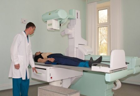
How is fluoroscopy of the esophagus and stomach performed?
The entire procedure of fluoroscopy with contrast of the esophagus and stomach is carried out in several stages:
- A survey x-ray is performed.
- The patient is given a sip of liquid barium contrast to drink. The entire act of swallowing and the movement of the suspension into the stomach is monitored on the screen.
- The esophagus is tightly filled with contrast. The condition of the organ and pathological changes in it are examined. Several x-rays are taken (frontal projection, two lateral, left oblique).
- If a study is planned in the Trendelenburg position, the patient is transferred to the required position and pictures are taken.
Next, they move on to examining the stomach. Pictures are taken both in a standing and lying position. If necessary, the intestines are further examined.
X-ray examinations are contraindicated for children, but for newborns, if there are signs of congenital pathology of the esophagus that require urgent surgery, X-rays with barium contrast are taken.
Survey results
The main benefit of a barium X-ray of the stomach is the ability to obtain data on the condition of the internal organs, in particular their structure and shape.
This helps to identify at different stages the presence of many pathologies, including cancer of the stomach and esophagus.
Shots from different angles give different results. For example, with an x-ray taken in a direct projection, you can see the relief of the esophagus and the antrum of the stomach, as well as assess the condition of the beards and folds of the upper section of the stomach and its body, and how normally the gastro-rectal junction functions.
This image will also show the structure of the esophagus and the folds of the duodenum. Most results can be obtained after the first dose of barium solution.
X-rays show the pneumorelief only after the patient takes half a glass of contrast barium.
Images with lateral and oblique projections are no less informative: with their help, you can determine whether there is displacement of the stomach, evaluate the relief of the antrum, as well as gastroesophageal reflux and the functionality of the pylorus.
Video:
An image taken from the Trendelenburg position will give the doctor information about the presence of a hernia in the esophageal opening, as well as about the condition of the muscle fibers - if they are weakened, the stomach may prolapse into the esophageal opening of the diaphragm.
Using pictures taken from the back, you can evaluate the elasticity and size of the organ.
Thus, the X-ray result mainly shows changes in the structure and shape of the stomach and esophagus.
Various changes in these values may indicate pathologies such as hiatal hernia, the presence of ulcers, polyps or cancerous tumors in the stomach or esophagus, reduction of the duodenal canals, impaired gastrointestinal motility, Ménétrier's disease, as well as varicose veins on the walls of the stomach or esophagus .
The complete examination takes approximately 40 minutes. In most cases, the examination result can be obtained almost immediately – within an hour.
However, in some cases, when it is not possible to immediately determine the pathology, the doctor may need more time - then the preparation of the results may take a day.
READ What diseases are diagnosed on an X-ray of the thoracic spine?