What is endosonography
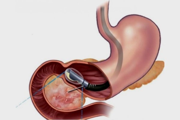
Endosonography of the stomach is considered a combined diagnostic method: an ultrasound sensor and an endoscope are used. The device is used as a guide for the sensor inside the digestive tract to its destination.
Endoultrasound is performed only in a hospital under the supervision of a physician. If the diagnostic and preparation rules are followed, errors in the results are excluded.
Endoscopic ultrasound is better than conventional ultrasound. During the procedure, ultrasonic waves are not scattered. Therefore, the structures of the stomach, pancreas and esophagus are completely visible.
Diagnostic value of ultrasound
Ultrasound of the abdominal cavity for abdominal pain is the main method of primary diagnosis of gastroenterological, urological, gynecological and surgical diseases. In many cases, the doctor only needs to look at the sonogram to understand what the patient is suffering from. However, in some cases, this study alone is not enough.
Ultrasound cannot determine the nature of free fluid in the abdominal cavity, find out the histological structure of the tumor, identify the cause of liver damage, and so on.
Moreover, with excessive gas formation, this research method becomes completely uninformative, and in the results the doctor receives the entry “pneumatosis intestinalis,” which has no diagnostic value.
Indications for the procedure
Indications for performing endosonography of the stomach are:
- Suspicions of the presence of malignant neoplasms in the stomach, esophagus and colon. Early diagnosis in most cases allows tumor removal using an endoscope. This is possible by detecting changes in the lymph nodes.
- Choice of treatment tactics. After performing an endosonography, a person, depending on the disease and stage, is prescribed endoscopy, laparoscopy or open surgery.
- Assessment of the condition of the organ after operations and treatment.
- Determination of the degree of deformation of the stomach wall in the presence of a sign.
- Finding out the causes of pain in the stomach if other diagnostic methods have failed.
How to do an ultrasound of the esophagus
An ultrasound can be done in several ways. The choice of technique depends on the expected disease and the individual characteristics of the patient.
- Transdermal. Mainly used in children. The doctor examines the esophageal tube by placing a probe on the neck, chest, and upper abdomen.
- Intraesophageal. The sensor is inserted into the lumen of the esophageal tube. This technique makes it possible to examine all layers of the organ. For use only in adults.
- Ultrasound with water-siphon test. It is used to examine the esophageal tube and stomach and assess the motor function of organs. Immediately before the procedure, the patient drinks 700-800 ml of water to fill the lumen. The inspection is carried out from the outside.
- Endoultrasound. It is a combination of endoscopy and ultrasound. The sensor is attached to the end of a flexible tube. The entire esophagus and stomach can be examined.
The most accurate and reliable are the intraesophageal method and endoUS. They are often accompanied by injury to the esophageal wall.
Sometimes for reliability it is necessary to do several research methods.
How to prepare for research
Correctly performing endosonography of the stomach and careful preparation make it possible to create an accurate picture of the condition of the organ. First, the doctor interviews the person and makes sure that he has no contraindications to EUS (endoscopic ultrasound sonography).
Before an endoscopic ultrasound examination, a person undergoes urine and blood tests to determine acute infections and diseases of internal organs. Usually such diseases are asymptomatic.
Preparation stages:
- A week before diagnosis, foods that contribute to bloating are excluded from the diet: cabbage, flour, legumes.
- 8 hours before the procedure, it is forbidden to eat or drink any liquid, as vomiting sometimes occurs during endosonography.
- On the day of the examination, do not smoke to prevent excessive salivation.
Ultrasound of the stomach and intestines - is it effective?
Ultrasound diagnostics is a very common research method these days. It is available in such industries as urology, cardiology, traumatology, gastroenterology, etc.
In the latter, parenchymal organs such as the liver, kidneys, pancreas, spleen and others are most often studied. However, sometimes the question arises: “Do they do ultrasound of the stomach and intestines?”
Yes, such research is performed, but much less frequently than in other areas of medicine.
What useful features does ultrasound have?
Let's start with one of the main advantages of ultrasound examination - safety. It is, for example, due to the properties of ultrasonic waves, which by their nature are loud sound waves inaudible to the human ear.
In nature, some animals use such waves for hunting (bats), communication (dolphins) and orientation in space.
Also, safety allows you to perform ultrasound as many times as necessary and, thus, monitor the dynamics of treatment or the development of the disease.
Another positive point is painlessness. Ultrasound of the stomach does not require skin punctures and does not cause pain. Therefore, this study can be carried out even in children.
What can you study with ultrasound of the stomach and intestines?
Ultrasound is the worst way to study the small intestine, since its loops overlap each other very much and, thereby, distort the results. However, the functioning of the small intestine can be studied, although not completely.
From the entire gastrointestinal tract you can examine:
However, it is immediately worth noting that ultrasound is rarely used to diagnose diseases of the gastrointestinal tract. This is due to the fact that these organs are hollow and have anatomical features that make them quite weakly susceptible to ultrasonic waves.
The following can be examined relatively well by ultrasound of the stomach and intestines:
- lesser and greater curvature of the stomach;
- the pyloric canal and cave (sphincter that separates the pyloric part of the stomach from the duodenum);
- the very zone of connection with the duodenum.
However, ultrasound of the stomach, most often, is either an additional research method, allowing a more detailed study of the pathology, or as the first stage of diagnosis, when most other methods are impossible or are not necessary.
What is better? Ultrasound of the stomach or FGDS
- Ultrasound of the stomach and intestines has some advantages. During an ultrasound, it is impossible to infect the examined organs, since the sensor only comes into contact with the skin and makes it possible to determine whether there are infiltrated oncological processes.
- But for example, on the side of FGD of the stomach, there is the opportunity to perform a biopsy, study the organ from the inside in more detail, evaluate the folds, determine the degree and nature of changes, etc.
However, it cannot be said that one method is better than another. It would be more accurate to note that they have a different direction and range of diagnosed pathologies.
Thus, ultrasound of the stomach copes well with the diagnosis of gastroesophageal reflux, cancer, disorders of evacuation function, and some inflammatory lesions.
However, a gastroenterologist must decide what will be best in a particular situation.
Indications for ultrasound of the stomach and intestines
If you have symptoms characteristic of the gastrointestinal tract, you should first consult a doctor, and only then go for a diagnosis. An ultrasound of the stomach and intestines may be performed if the following diseases are suspected:
- inflammatory pathologies of the stomach and intestines;
- benign and malignant neoplasms;
- calculous cholecystitis
- if there are liver pathologies;
- achalasia of the esophagus;
- gastroesophageal reflux;
- yellowness of the skin and sclera;
- B12 deficiency and folate deficiency anemia;
- varicose veins of the esophagus and stomach.
Preparation for ultrasound diagnostics of the stomach
- It is not difficult to prepare for an ultrasound of the stomach. You need to follow a diet 3 days before the test that excludes all gas-forming foods from your diet. Among them are: bananas, cabbage (as well as many other raw vegetables and fruits), fermented milk products, fresh baked goods, black bread, etc. The diet is simple and does not require much effort.
- 8 hours before the scheduled time of the study, the patient should not eat, because this can also provoke the formation of excess air in the intestinal lumen, which will greatly distort the results. For this reason, most often, the examination is scheduled in the morning.
- The duration of fasting, if the examination is not in the morning on an empty stomach, should be about 8 hours. If the patient suffers from severe hunger pains, then the duration of no food intake is reduced to 4 hours. On the eve of the study, the last meal should be no later than 8 pm.
In preparation for an ultrasound of the stomach and intestines, there is also a place for medications. If preparation is violated, you should inform your doctor. If the diagnosis is not tolerated, then most likely the patient will be prescribed carminatives.
The same, for example, applies to patients suffering from flatulence. Simple preparation is another advantage of this diagnosis.
How is an ultrasound of the stomach done?
- The subject comes to the diagnostic room at the appointed time and lies down on the couch, exposing the anterior abdominal wall.
- Then, a special gel is applied to the examination area.
It is hypoallergenic and is necessary to remove excess air between the skin and the sensor. - After this, the doctor proceeds directly to the ultrasound examination of the stomach itself, during which he examines the organ in various projections.
Applying Contrast
When examining the stomach using ultrasound, contrast is also used. The contrast agent for ultrasound examination is water. A small amount of liquid (300-350 ml), drunk in small sips, in order to avoid swallowing air, will make it possible to study the walls of the stomach in more detail.
Results and norms of ultrasound of the stomach and intestines
The initial assessment is carried out by a sonologist, directly during diagnosis and then when comparing the obtained data with the norms more fully. All results should then be reviewed by a gastroenterologist. If you do not introduce the specifics of ultrasound examination, then ultrasound of the stomach evaluates the following:
- wall thickness (depending on the department) varies from 4-6 mm to 6-8 mm ;
- the wall consists of clearly differentiated 5 layers ;
- the thickness of the submucosa is about 3 mm ;
- the mucosa is about 1.5 mm ;
- the lamina propria of the gastric mucosa is 1 mm ;
- a glass of liquid drunk before the study is evacuated from the stomach in 20-25;
- uniformity of layers of the gastric wall.
However, the indicators listed above are not all the data that is assessed during diagnosis. The echogenicity of certain gastric structures on ultrasound plays a very important role.
Echogenicity is the ability to reflect ultrasonic waves. For example, the submucosa has medium echogenicity, and the muscularis propria has low echogenicity.
What does an ultrasound of the stomach show?
An ultrasound of the stomach can diagnose various pathologies. They have varying levels of danger to life, but certainly all these pathologies cause trouble, because stomach diseases rarely occur without painful symptoms.
- Gastroesophageal reflux disease (GERD). This pathology has already been mentioned above, however, what is hidden behind such a complex name. This pathology is characterized by the reflux of the contents of the stomach or duodenum into the esophagus. Symptoms that arise in this case: heartburn;
- sour belching;
- pain behind the sternum, which spreads to the interscapular space, neck and even lower jaw (therefore, a careful differential diagnosis between GERD, cardiac and pulmonary pathology is needed).
These symptoms usually appear after eating, at night, or when bending forward. Many people experience these very unpleasant sensations. However, the danger of this disease lies in permanent damage to the lower esophagus.
On an ultrasound of the stomach, this disease can be noticed during functional tests, when the patient is asked to change his body position. If previously drunk liquid is thrown into the stomach, the doctor will definitely register this using an ultrasound machine.
Treatment of this disease is simple and consists of searching for the cause of GERD, as well as drug therapy.
- Pyloric stenosis. But this pathology is much more dangerous than GERD and occurs mainly in children 2-3 weeks of age or, less often, in patients who, for some reason, have scars in the area of the pyloric sphincter (pylorus). The pathology itself is characterized by a narrowing of the pylorus to a critical level, when food and even liquid cannot pass further. Symptoms of this pathology in children are as follows:
- vomiting like a fountain of just eaten food;
- children are developmentally delayed;
- signs of dehydration appear and progress rapidly;
- constipation and scanty stool;
- decrease in the amount of urine.
in children, the amount of milk in the vomit will be more than what he ate, and this milk will also be curdled;
In adults, the symptoms are slightly different:
- constant heaviness and nausea after eating;
- the pain is bursting;
- after bouts of vomiting, the patient feels relief;
- constant belching;
- weight loss, decrease in the amount of vitamins and minerals in the body.
However, looking at the symptoms, even a person without medical education will understand that the mechanism of development of these diseases is almost identical.
For example, in children and adults, during an ultrasound of the stomach, the doctor will detect pyloric stenosis. Hypertrophic pyloric stenosis is most easily detected when the cause is overgrown muscle tissue. However, ultrasound can detect other types of this disease.
Differentiation between pyloric stenosis and pylorospasm is very important. Since the latter is not so dangerous and its treatment is much simpler.
If you perform a timely ultrasound of the stomach of a child with pyloric stenosis, this will help avoid more severe consequences for this pathology, and in advanced cases it can save a life.
Treatment of pyloric stenosis is only surgical, during which the tissue is dissected and the pyloric lumen is increased.
Ultrasound for gastric ulcers is rarely used, since preference is given to fibrogastroduodenoscopy. However, ultrasound is increasingly being used to identify peptic ulcers. Its accuracy in this case is 85-90%.
Reviews from doctors and patients
Ultrasound of the stomach and intestines, although not used often enough, takes its place of honor among methods for diagnosing diseases of these organs.
Patients highlight the ease of diagnosis, its speed, simple preparation, painlessness and safety. Doctors also note that ultrasound helps establish a number of diagnoses without the risk of exposing the body to harmful rays.
This is especially true for diagnosis in children, as well as situations where the clinical manifestations of the disease are very vague.
Ultrasound is a safe and accessible diagnostic method that works well, but we should not forget about many other types of examination.
SHARE WITH YOUR FRIENDS AND VOTE: ( 3 5.00 out of 5) Loading…
Source: https://uzipro.ru/uzi-zheludochno-kishechnyiy-trakt/uzi-zheludka.html
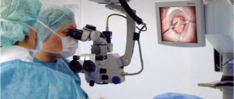
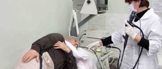
How is endosonography of the stomach done?
Endoultrasound of the stomach involves inserting a probe through the oral cavity. Sometimes the diagnosis is carried out under anesthesia or painkillers so that the person does not feel pain.
When neoplasms are detected, a biopsy of the organ tissue and adjacent lymph nodes is performed.
After EUS, unpleasant symptoms may appear: headaches, nausea, vomiting and flatulence. The discomfort listed above is temporary.
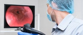
Watch a video of how EUS is performed:
Decoding the results
In a normal state, when performing endoscopic ultrasonography, the structure of the gastric walls is clearly distinguishable by echogenicity, the ability of internal organs to reflect ultrasound waves:
- the thin surface of the mucosa is hyperechoic;
- increased echogenicity of the submucosal layer, it is wider on the device screen;
- increased or moderate echogenicity of the serous and outer adventitia;
- the muscle layer appears with little echogenicity.
If the organ is healthy, the thickness of its walls varies between 5–6 centimeters in the pyloric region. The tissue structure is not disturbed and there is no irritation.
The layers of stomach tissue have a certain thickness throughout the entire wall. With the development of interstitial tumors, rounded dark formations with an uneven thickened wall with different echogenicity are visible on the screen:
- on EUS, the lipoma appears as a hyperechoic round area;
- Abrikosov's tumor looks like a homogeneous mass with a vague contour;
- gastrointerstitial tumors are shown in the form of dark circles with reduced density, sometimes with anechoic, hyperechoic inclusions;
- leiomyoma is visible as a homogeneous zone with a clear contour and reduced density.
In the results, the sonographer indicates the size of the tumors, location, echogenicity and the layer of the stomach wall from which they developed. In addition, the condition of the regional lymph nodes is deciphered.
Ultrasound endoscopy shows the presence of a stomach ulcer with changes in the thickness of the walls of the organ.
In the video, watch a case of diagnosing a tumor using EUS:
Thickening of the stomach wall on ultrasound: what are the causes, diagnostic features
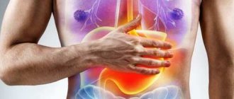
In case of stomach diseases, one of the most informative and safe examinations for the patient is ultrasound diagnostics. When performing it, you can study the structure of the organ and peristalsis in real time. This is a hollow muscular organ and a sign such as thickening of the stomach wall indicates pathological changes in it.
Thickening of the stomach wall
The stomach performs several important functions in the body. It breaks down food products under the influence of hydrochloric acid and enzymes that are produced by glands located in the thickness of the mucosa. In addition, under the action of peristalsis - muscle contractions - food masses in the stomach are mixed, crushed, and moved into the intestines.
It also performs a protective function, disinfects food, destroying pathogenic microbes that come with it, and protects the abdominal cavity from the aggressive action of gastric juice.
All this is ensured by the special structure of the walls of the organ, therefore, if any changes are detected in them on ultrasound, this may give the diagnostician a reason to suspect the development of a number of diseases, including such a dangerous pathology as cancer. Let's consider what thickening of the walls of the stomach indicates, what it is, and what diagnostic value does this indicator have?
What should be the normal thickness of the stomach wall?
The stomach wall consists of three main layers, each of which has a specific function. They are clearly distinguishable during ultrasound examination:
- Slimy . Contains glands that produce hydrochloric acid, pepsinogen and other enzymes. On ultrasound it appears hypoechoic (dark line). It contains the mucosa itself and the submucosa, in which glands and blood vessels are located.
- Muscular . Consists of several rows of multidirectional muscle fibers. Their contraction provides peristalsis. Appears as a hyperechoic thin line.
- Serous . The subserous and serous layers are also distinguished; their density on ultrasound relative to the muscular layer is low, that is, the serous membrane looks hypoechoic.
What does thickening of the stomach walls mean?
When examining the stomach, the diagnostician pays attention not only to the thickness of the walls and its changes in different parts, but also to their rigidity, lack of peristalsis in a certain area, and smoothing of the relief of the folds. The detection of such signs may be the first symptom of a developing pathology, even with normal thickness.
The physiological causes of thickening of the stomach wall on ultrasound are:
- inflammatory reaction causing tissue infiltration and swelling;
- benign, malignant neoplasms with exophytic growth into the gastric cavity at an early stage;
- interstitial tumors growing along the muscle layer.
Causes of thickening of the stomach walls
The main reasons leading to thickening of the stomach walls are as follows:
- gastritis, in which swelling of the mucous membrane and hypertrophy occur as a result of the inflammatory process;
- traumatic injuries cause inflammation with subsequent sclerosis of the damaged area;
- peptic ulcer, thickening caused by the formation of connective tissue (scar) around the lesion;
- Ménétrier's disease, a chronic disease characterized by hypertrophy of the mucosa with the formation of cysts and adenomas;
- benign formations (polyps), at an early stage they slightly rise above the mucous membrane, causing a feeling of thickening of the wall;
- malignant tumors of the stomach (adenocarcinoma, sarcoma, rhabdomyoma);
- tissue infiltration during specific infections (syphilis, tuberculosis, actinomycosis).
Changes that occur in the wall of the stomach and are detected on ultrasound are called hollow organ syndrome.
What are the benefits of undergoing an ultrasound of the stomach can be found in this video.
Ultrasound diagnosis of thickening of the stomach walls
Wall thickening can also be detected using x-rays, but ultrasound has several advantages. The examination allows you to study the organ in different projections, it is safe and does not have a harmful effect on the body. During the examination, the Doctor can see changes in each layer.
On ultrasound, the wall looks like a thin line consisting of three layers - hyperechoic inside and hypoechoic on the sides. The mucous membrane has folds that smooth out when the sensor is pressed. Normally, movements (peristalsis) can also be observed.
An increase in thickness occurs due to any of the layers. Thus, with interstitial tumors, changes in the size of the submucosal or muscular layer in a certain area are often detected. An increase in the mucous layer occurs during inflammatory processes and hypertrophy.
Also, benign (polyps) and malignant (adenocarcinoma) neoplasms often form in this layer. Doppler examination allows one to differentiate malignant neoplasms, which can detect increased blood flow in this area.
Source: https://uzi.guru/zhivot/zhl/diagzh/utolshhenie-stenki-zheludka.html
Contraindications
Ultrasound endoscopy of the stomach, in contrast to the FGS procedure, has fewer contraindications. In general, diagnosis is not carried out among people whose esophagus is blocked. Common causes of the symptom are stenosis and achalasia. Because of this, the endoscope does not pass through the wall of the stomach and esophagus.
EndoUS is prohibited for decompensated diseases of internal organs, disorders of the cardiovascular and respiratory systems. The procedure is excluded during the period of exacerbation of chronic forms of gastrointestinal diseases.
Combined diagnostics using an endoscope and an ultrasound sensor is done not only to determine the presence of diseases. Sometimes the study is used for therapeutic purposes, when sclerotherapy is necessary in the presence of dilated vessels of the gastrointestinal tract.
Advantages and disadvantages of endoscopy with ultrasound
EndoUS is a safe and informative method that allows you to identify changes in deep-lying internal organs. Unlike gastroscopy, endosonography shows the layer-by-layer structure of the stomach wall, interstitial neoplasms and regional lymph nodes. Depending on the sensitivity of the equipment and the qualifications of the specialist, tumors 1 mm in diameter can be detected.
Computed tomography and magnetic resonance imaging can also show small changes. But EUS helps to evaluate the picture as a whole, and with layer-by-layer tomography, small elements may not fall into the scanning area. In addition, CT is an x-ray method that carries radiation exposure.
How much does diagnostics cost?
The cost of gastric endosonography in 2020 varies between 1,500–11,000 rubles. The price depends on the region and the classification of the sonographer. Thus, in the Moscow region, endosonography costs 3,000–10,000 rubles on average. In St. Petersburg the price is much higher – 4,000–11,000 rubles.
Endosonography of the stomach is considered a modern diagnostic procedure, which, when properly performed and prepared, gives the most accurate results of the condition of the organ.
Have you undergone such research? Tell us about it in the comments. Share the article with your friends on social networks. Be healthy.
Hypertrophic gastritis: causes, signs, symptoms and treatment
If the mucous membrane grows in the gastrointestinal tract, hypertrophic gastritis occurs. The disease is accompanied by pain in the epigastric region, heartburn, and lack of appetite. At the first signs of deterioration in health, it is recommended to consult a doctor who will diagnose, prescribe the correct treatment and give preventive recommendations.
How and why does pathology occur?
Adenomatous formations that occur with hypertrophic gastritis can degenerate into malignant ones.
Under the influence of unfavorable factors, the mucous tissue of the gastric epithelium grows, thickens and becomes deformed. Cysts and polyps, as well as benign tumors - adenomas, form on the thickenings. The pathological process is preceded by the following factors:
- bad habits;
- non-compliance with food intake;
- improper diet;
- regular stress;
- uncontrolled medication intake;
- reflux, in which food from the small intestine is thrown into the stomach;
- lupus erythematosus;
- violation of the acidity level of gastric juice;
- food allergies;
- deterioration of digestion caused by damage to the intestinal villi;
- fungal and bacterial infections, including Helicobacter pylori.
Reasons for development
Adequate and correct treatment of hypertrophy can only be done if the cause of the disease is correctly established. The disease in most cases develops in a similar way to other forms of gastritis. However, this type of disease has its own individual characteristics and characteristics that fundamentally distinguish it from other types of inflammatory processes in the gastrointestinal tract.
The occurrence of the disease may be associated with the following factors:
- Infection with Helicobacter pylori is the main cause predisposing to the development of a pathological process in the stomach. This microorganism enters the human body along with poor quality and poorly processed food.
- Violation of the normal diet - a complete diet, which contains a sufficient amount of fiber, proteins, carbohydrates - is practically a guarantee of the absence of disease. However, frequent overeating or undereating, a poor and monotonous diet lead to the rapid appearance of an inflammatory process.
- Chronic hyperacidity of the stomach - an excessively acidic environment of the stomach predisposes to more active proliferation of pathogenic bacteria and microorganisms. Frequent stress, frequent use of spices and hot sauces, coffee, strong tea, and some medications predispose to increased acidity.
- The presence of Reflux - in this condition, a person experiences an involuntary reflux of stomach contents into the esophagus, which causes a lot of unpleasant sensations and at the same time negatively affects the condition of the digestive tract as a whole. The development of such a condition is predisposed by a person’s constant nervousness, stress, depression, and disruption of the autonomic nervous system.
Important: With hypertrophy, self-medication does not bring the expected results. The disease can reduce the intensity of its symptoms only for a short period of time. Then the symptoms resume with a vengeance.
Classification of gastropathy
Doctors distinguish the following types of pathology shown in the table:
| Form of the disease | Peculiarities |
| Hypertrophic granular gastritis | Formation of cysts and polyps |
| Warty | The appearance of warts on the mucous membranes of the stomach |
| Erosive | The appearance of ulcers on the stomach linings |
| In the antrum | The presence of nodules, erosion and compaction in the antral folds |
| Giant (Menetrier's disease) | Formation of large folds |
| Unnatural thickening on the gastric mucosa | |
| Diffuse | Gland atrophy |
| Inflammation of epithelial tissue | |
| Death of stomach cells | |
| Obstructive | Inflammation spreads throughout the organ |