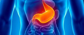
Dilatation of the esophagus
– cylindrical or spindle-shaped increase in the lumen of the esophagus (diffuse or local) with impaired evacuation of food into the stomach. Clinically manifested by dysphagia, chest pain, regurgitation of food into the oral cavity, emaciation, and night cough. To make a diagnosis, esophagoscopy, radiography of the esophagus, and esophageal manometry are performed; according to indications of ultrasound or MSCT of the abdominal organs, scintigraphy of the esophagus. Treatment is aimed at eliminating the cause of the disease; surgical intervention may be required (balloon dilatation of the cardiac sphincter, excision of esophageal diverticulum, resection of the esophagus for cancer).
Dilatation of the esophagus
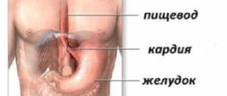
Dilation of the esophagus is a fairly rare condition that occurs against the background of another pathology. The most significant cause of diffuse dilatation of the esophagus is achalasia cardia. Much less often, diverticula, inflammatory and adhesive processes in the mediastinum lead to an increase in the lumen of the esophagus. In some patients, it is not possible to establish the exact cause of the development of this pathology. The formation of dilation of the esophagus is based on the difficulty of evacuating food masses into the stomach due to an obstacle, usually located in the lower parts of the esophagus or at the entrance to the stomach (cardiospasm, achalasia of the cardia; cancer of the esophagus; adhesions that spread across the esophagus and pull it over). Gradually accumulating food masses stretch the walls of the esophagus, disturbances in its motility occur, and organic changes are formed in the tissues with persistent deformation of the esophagus.
Causes of esophageal dilation
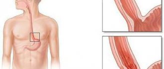
Dilatation of the esophagus is formed against the background of cardiospasm, achalasia cardia, and esophageal cancer; inflammatory processes in the mediastinum, leading to scarring and the formation of traction diverticula; adhesions that stretch the lumen of the esophagus. Based on the mechanism of occurrence of esophageal dilation, the following forms of the disease are distinguished: diffuse dilation (cylindrical, spindle-shaped, S-shaped deformation with esophageal dilatation) and local (esophageal diverticula). The general mechanism for the formation of diffuse expansion of the esophagus is the presence of an obstacle to the passage of food into the stomach with gradual stretching of the walls of the esophagus by accumulating food masses.
Most often, dilatation of the esophagus is diagnosed in patients suffering from cardiospasm or achalasia cardia. These two conditions are stages of one disease, in which at the beginning a functional disorder is formed in the form of a transient spasm of the lower esophageal sphincter, and as a result of the progression of pathological processes in the distal parts of the esophageal tube, organic changes begin with the development of constant achalasia (lack of relaxation) of the cardia.
In the pathogenesis of esophageal dilatation with achalasia cardia, three mechanisms are distinguished: disruption of the autonomic regulation of relaxation of the cardiac sphincter, frenospasm and achalasia cardia itself. Autonomic disorders can occur against the background of severe emotional shocks, leading to changes in the tone and motility of the esophagus, and a failure of the mechanism of opening and closing the cardia. In addition, disturbances in the autonomic regulation of the esophagus can occur against the background of other pathologies of the abdominal organs (urolithiasis and cholelithiasis, pancreatitis, chronic gastritis, gastric and duodenal ulcers, liver tumors, etc.) according to the type of visceral reflexes.
The complex mechanism for evacuating food from the esophagus to the stomach also includes contraction of the muscle fibers of the diaphragm. Research in the field of gastroenterology has shown that peristalsis of the esophagus begins in its upper parts and ends in front of the esophageal opening of the diaphragm. After this, the diaphragmatic shutter is activated, pushing the food bolus into the stomach. Spasm of the muscle fibers of the diaphragm (phrenospasm) can lead to blockage of the lumen of the esophagus and its subsequent expansion.
Hemorrhoids kill the patient in 79% of cases
The combination of functional cardiospasm against the background of autonomic dysfunction and frenospasm sooner or later leads to the formation of organic scar changes in the distal parts of the esophagus and cardiac sphincter with the development of achalasia cardia. The passage of food masses through the lower sphincter of the esophagus is significantly hampered; they accumulate in the lumen of the esophagus, stretching its walls. The muscle tone of the esophagus gradually decreases, which first leads to an expansion of its lumen, and in the future an S-shaped deformation is formed due to the elongation of the esophageal tube. At this stage of the disease, an enlargement of the upper parts of the esophagus occurs, and regurgitation of food and liquid into the oral cavity occurs, especially pronounced in an upright position. The entry of food masses into the respiratory tract during regurgitation leads to the development of bronchitis and pneumonia. Congestion in the esophagus also ends in an inflammatory process (esophagitis) and the appearance of an esophageal ulcer.
Symptoms of esophageal dilatation
The clinic of esophageal dilatation is developing gradually. At the beginning, the symptoms are transient, but over time, against the background of organic changes in the esophagus, the intensity of the symptoms increases, concomitant diseases and complications develop, which, if left untreated, can lead to the death of the patient.
In the initial stages of the disease, the patient is bothered by dysphagia and chest pain. In the presence of cardiospasm, the first manifestations may be sudden: against the background of fear or severe emotional shock, there is a feeling of a lump in the throat, pain in the area of the xiphoid process or behind the sternum. These symptoms soon disappear, but reappear after some time. Gradually, episodes of dysphagia become more frequent and no longer go away on their own. To improve the movement of food into the stomach, the patient can make certain efforts: compress the lower parts of the chest, constantly wash down food with water, swallow air, and so on. The pain behind the sternum also intensifies, radiating to the epigastrium, shoulder blades, and left arm (may resemble the pain of angina pectoris). A feeling of fear before eating is formed.
The symptoms of obstruction increase due to the expansion of the esophagus. The patient often complains of intractable attacks of hiccups and regurgitation of eaten food. Occasionally, profuse vomiting of undigested food masses without the admixture of hydrochloric acid and bile occurs, which brings significant relief, sometimes vomiting even leads to a temporary disappearance of symptoms.
Due to the fact that the esophagus is constantly overcrowded, the expansion extends to its upper sections, which is why at night, in a horizontal position, liquid food masses flow out and enter the vocal cords and respiratory tract. A symptom pathognomonic for dilatation of the esophagus appears - night cough. Bronchitis develops, and then aspiration pneumonia and bronchiectasis. Due to the fact that food practically does not enter the stomach, and patients are often forced to induce vomiting to alleviate the condition, exhaustion develops, which, in combination with concomitant serious diseases, can even lead to the death of the patient.
Enlargement of the esophagus should be differentiated from gastroesophageal reflux, mediastinal tumor, bronchiectasis, tuberculosis, coronary heart disease, neurogenic dysphagia, damage to the esophagus due to amyloidosis and scleroderma.
How to distinguish esophageal diseases from heart problems?
In case of heart disease, heartburn, bitter or sour belching, and increased salivation do not occur. A person without appropriate education may confuse chest pain caused by angina with spasm due to esophagitis.
| Symptoms | Heart diseases | Problems with the esophagus |
| Differences | ||
| Salivation | Absent | Present |
| Heartburn | Absent | Present |
| Difficulty swallowing | Absent | Present |
| Pain | Pulling, pressing, radiating to the left jaw, shoulder blade, shoulder or arm. | Burning, sharp, passes into the throat or stomach. |
To exclude pain of cardiac origin, the patient is prescribed an ECG.
Problems with the esophagus significantly reduce a person’s quality of life. Pain and dysphagia are considered the main symptoms of the development of pathologies. It is recommended to visit your doctor regularly in order to diagnose the disease in a timely manner.
Diagnosis of esophageal dilatation
When the first symptoms of esophageal dilatation appear, you should consult a gastroenterologist. Upon examination and examination of the patient, an expansion of the boundaries of the dullness above the mediastinum is revealed; sometimes a soft elastic protrusion on the left neck, containing food masses and liquid, is palpated.
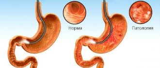
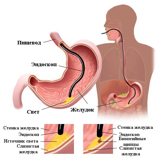
The most informative methods for diagnosing enlarged esophagus are examination by an endoscopist with esophagoscopy and radiography of the esophagus. Esophagoscopy is possible only after the evacuation of liquid masses from its lumen - the phenomena of esophagitis and ulceration are visualized. Using this study, it is possible to identify the cause of the dilation of the esophagus (achalasia cardia, tumor, scars and adhesive constrictions, diverticula).
On X-ray with contrast, the lumen of the esophagus is expanded and filled with food masses. The contrast agent settles for a long time, in the form of snow flakes. Evacuation of contrast from the esophagus is significantly delayed (more than several hours). Esophageal manometry allows you to identify esophageal motility disorders. For differential diagnosis, ultrasound and MSCT of the abdominal organs, scintigraphy of the esophagus are performed.
Treatment with traditional methods
One of the conservative ways to relieve reflux esophagitis is treatment with folk remedies. They use herbs and infusions that have an astringent effect.
- A tablespoon of oak bark is combined with cinquefoil rhizomes, 40 g of dry walnut leaves and 20 g of oregano. Mix with three cups of unboiled water, leave for 3 hours, then boil for 5 minutes. Cool and take a quarter of a mug before meals.
- Mix 30 g of oregano, 20 g of alder, 40 g of violet flowers, 10 g of burnet from the table and pour in 300 ml of boiling water. Insist, take 3 times a day.
- Dandelion flowers are placed in the bottle, sprinkling each layer with sugar. Pound until syrup is obtained and stand for 6 hours. 10 g of infusion is diluted with 100 ml of water and taken once a day.
- Sea buckthorn oil: take a teaspoon half an hour before meals.
Treatment with folk remedies for reflux esophagitis is possible only after consultation with a doctor as an additional remedy to the main method.
Treatment and prognosis of esophageal dilatation
The main focus of treatment for dilated esophagus is to eliminate the cause of this condition. If dilatation of the esophagus has formed against the background of achalasia cardia, the patient should be explained the importance of following a daily routine and diet. The psychological state of the patient is of great importance for restoring normal autonomic regulation and eliminating frenospasm, therefore the task of the attending physician is to reassure the patient and instill in him faith in a successful outcome of the disease.
A special diet and anti-inflammatory treatment are prescribed. Food should be gentle chemically, mechanically and thermally. To avoid stagnation, the esophagus should be emptied of its contents before bedtime. It is recommended to drink alkaline waters and herbal decoctions to rinse the esophagus. Medications include vagosympathetic blockades, B vitamins, and antispasmodics.
If conservative therapy is ineffective, balloon dilatation of the cardiac sphincter and bougienage of the esophagus are performed to restore its patency. Balloon dilatation is contraindicated against the background of esophagitis, fissures and ulcers in the area of stenosis, as it can lead to rupture of the esophagus. With significant organic changes in the cardiac sphincter, cardiomyotomy surgery may be required. In weakened patients and in the presence of contraindications to surgical restoration of esophageal patency, gastrostomy can be performed until the condition stabilizes. If diverticula are present, they are excised. If a patient is diagnosed with esophageal cancer in the early stages, resection of the esophagus is performed, followed by plastic surgery.
The prognosis for dilatation of the esophagus is favorable, but the earlier it is started, the higher the effectiveness of treatment (in the early stages of the disease, the effectiveness of surgical intervention is more than 90%). Specific prevention of esophageal dilatation has not been developed. Diseases that can lead to this condition should be promptly identified and treated.
Basic diagnostic techniques
The appearance of pain in the larynx can have several causes, and diseases of the esophagus are one of them. Their presence is accompanied by pronounced symptoms and can cause serious discomfort to a person.
Pain in the throat, problems with swallowing and sensations of a foreign body in the throat are just a small part of the symptoms that can be observed with pathological damage to the esophagus. The appearance of such symptoms serves as a signal to immediately consult a doctor, and ignoring them, on the contrary, can significantly aggravate the situation and lead to various complications.
A timely visit to a gastroenterologist makes it possible to determine the source of the problem and neutralize it, and the following techniques will help to do this:
- endoscopy of the esophagus;
- tissue biopsy;
- radiography;
- spiral tomography;
- pH-metry of the esophagus;
- endoscopic sound analysis.
The above methods make it possible to obtain the most complete and detailed answer to the question of how to check the esophagus. Moreover, each of them has its own characteristics and structure, and their effectiveness is at a fairly high level.