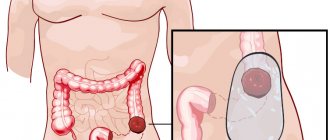
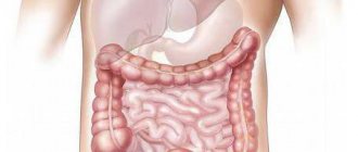
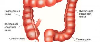
Neoplasm in the intestine
A tumor of the rectum, the symptoms of which are often similar to other diseases in this area, according to WHO, ranks 3rd among malignant neoplasms of the gastrointestinal tract and 4th among all oncological pathologies.
Rectal neoplasms are divided into benign and malignant. The latter are extremely dangerous. In this case, a tumor is formed due to a mutation in the cells lining the mucous membrane of the organ.
Malignant formation in this area is characterized by rapid development and invasion into nearby and distant organs. Rectal tumors have symptoms that are vague at first, and are characterized by frequent relapses, even with qualified treatment.
Classification
Oncology of the rectal area includes, depending on the histological structure, the following subtypes:
Types of disease
- Adenocarcinoma. The most common type, accounting for approximately 75% - 80% of all cancers in this area. The formation is based on glandular tissue. It mainly develops in people over 50 years of age. Cells are examined to determine the degree of differentiation (highly, moderately, poorly differentiated cells). A lower grade has a negative prognosis.
- Signet ring cell. A rarer form, the prevalence is approximately 3% - 4%. The name comes from the appearance of the cells observed under a microscope: a lumen in the central part with a rim and a cell nucleus at the periphery. This type of rectal carcinoma has a poor prognosis, with most patients not surviving more than 3 years.
- Solid cancer. An infrequent variety, consisting of glandular tissue, has a low degree of differentiation of cells arranged in the form of layers.
- Skirous (skyr). Another not common species. Characterized by a large amount of intercellular substance with a small number of cells.
- Squamous. In terms of frequency of occurrence, this malignant neoplasm of rectal cells is in third place (2-5%). An aggressive tumor that metastasizes quite early. It is believed that it develops due to infection with human papillomavirus infections. In almost all cases it is located in the anal canal area.
- Melanoma. A tumor consisting of melanocytes (known for the fact that when located on the skin it resembles a mole). One of the most dangerous, it metastasizes quickly, is rarely diagnosed at an early stage, and is difficult to treat.
Rectal cancer is divided depending on the direction of growth:
- Exophytic cancer. Growth is observed towards the inside of the organ.
- Endophytic. The tumor grows into the wall of the organ.
- Mixed form. It grows in both directions.
There is also a benign form of tumors (among which formations tend to often become malignant), which has its own classification:
- epithelial;
- carcinoid;
- non-epithelial.
The first category includes the following types of formations:
- Polyp.
- Villous tumor. It is part of the epithelium of the rectal area, is prone to malignancy, and is removed.
- Familial diffuse polyposis of the colon.
- Papilloma on the rectal mucosa.
- Carcinoid is a benign tumor that produces hormones (does not metastasize). The symptoms depend on the specific substance produced.
Prevention of colorectal cancer
The outcomes of colorectal cancer can be predicted based on the following data:
- Stages of the disease;
- Type and degree of tumor differentiation;
- Age and general condition of the patient;
- The presence of concomitant pathologies;
- Timeliness, adequacy and effectiveness of the treatment provided.
Depending on this, the prognosis for rectal cancer may be as follows:
- Cancer of the anal canal and lower ampullary rectum has the worst prognosis even at stages 1-2, as it requires disabling surgery and often recurs. Such patients are forced to use colostomy bags for life;
- Poorly differentiated tumors always have a much more favorable prognosis compared to tumors with a high degree of differentiation of cancer cells;
- Prognosis for life and recovery is significantly aggravated by old age, concomitant diseases and disorders of the general condition of patients. These factors not only limit the possibilities for performing radical surgery, but also accelerate the progression of the tumor process;
- The survival rate of patients in relatively satisfactory general condition with stage 1-2 cancer ranges from 60% to 85%;
- For stage 3 tumors in patients with relatively good health, survival for 5 years after diagnosis, subject to radical treatment, is about 30%;
- With stage 4 cancer, the prognosis for life is unfavorable. Almost all patients die within 6-8 months.
- Refusal of radical treatment of operable forms of cancer of any stage has an unfavorable prognosis and ends in death within a year.
Preventing colorectal cancer is not easy. This is due to the fact that it is not always possible to influence all its causes.
But it is within the power of every person to eradicate those risk factors, the presence of which increases the likelihood of developing this disease tenfold, or to do everything so that the disease that arises is detected as early as possible:
- Promptly treat chronic diseases of the rectum and anal canal (hemorrhoids, fissures, fistulas, etc.);
- Fight constipation;
- Avoid excess consumption of animal fats and fast food and enrich your diet with vegetable oils;
- Minimize contact with hazardous chemicals;
- Undergo preventive examinations once or twice a year.
Of course, all these measures do not guarantee 100% protection against colorectal cancer, but they significantly reduce the risk of its occurrence.
Author of the article: Bykov Evgeniy Pavlovich | Oncologist, surgeon
Education: completed residency at the Russian Scientific Oncology Center named after. N. N. Blokhin" and received a diploma in the specialty "Oncologist"
Other doctors
‹
15 scientifically proven beneficial properties of sesame!
How to fall asleep in 1-2 minutes?
›
Depending on the stage of anal cancer, surgical treatment can help achieve long-term remission: 5-year survival after surgery in the absence of regional metastases is 55-70%, and in their presence - about 20%. Combined chemoradiotherapy can cure anal cancer in 80% of patients with a tumor less than 3 cm in diameter. Relapses occur in less than 10% of cases. To assess the results of treatment for anal cancer and prevent recurrence of the disease, regular examinations by a proctologist are necessary.
Measures to prevent anal cancer and reduce the risk of HIV and HPV infection include the use of condoms during casual sex, having a regular sexual partner, quitting smoking, and regular screening examinations (PAP test and anoscopy).
Symptoms
At first, signs of the disease may be completely absent. It is almost impossible to detect cancer at an early stage, only during a routine examination or in the presence of other complaints.
As a benign and malignant tumor of the rectum grows, it also manifests itself with other signs:
- discomfort and pain during bowel movements;
- liquid or dense stool;
- blood or mucus may appear;
- malfunction.
Even during this period, the disease can easily be confused with hemorrhoids, which also manifests itself in the final stages as hemorrhages. A characteristic difference is the period of their appearance. Tumors in the rectum also have symptoms of an inflammatory process - pus and mucus.
There are also symptoms of a malignant tumor of the rectum, which are characteristic of different sexes. In women, tumor growth into the vaginal wall can lead to the formation of a fistula. Then, along with vaginal discharge, feces and pus appear.
In men, tumors often grow into the walls of the bladder. In this case, chronic cystitis develops and stool appears in the urine. Pathogenic organisms can penetrate further, causing pyelonephritis.
Diagnosis of pathology
Oncologists who treat colorectal cancer are guided by general principles.
The main principle is to make a correct diagnosis in a timely manner and prescribe appropriate treatment. At the same time, it is necessary to identify the reasons that caused the pathology.
It is important for the attending physician to know when the first symptoms of the disease appeared, which the patient himself noticed.
Depending on the duration of the disease, the pathology is divided into 4 stages. Various diagnostic methods are used to determine the stage.
Finger examination
Before starting to examine a patient for the presence of rectal cancer, the proctologist takes a detailed medical history.
This rule applies equally to men and women.
For a correct diagnosis, it is necessary to know about close relatives who suffered from diseases of the rectum.
Heredity plays a significant role in predisposition to cancer.
The causes of cancer are often determined by the genetic characteristics of the body.
The simplest and most effective method of primary diagnosis is considered to be digital examination.
Using this method, the proctologist has the opportunity to detect neoplasms in the rectum that are not characteristic of a healthy state of the body.
Video:
It is important to emphasize that the study only records the presence of tumors on the inner surface of the rectum.
It is impossible to determine the pathogenicity or benignity of a tumor using this study.
If a tumor is detected, the doctor prescribes additional testing to determine the causes of its appearance. It is important to obtain information about the presence or absence of cancer.
Instrumental methods
The causes of rectal cancer are identified using more effective methods than digital examination.
READ Folk remedies for the treatment of colorectal cancer
The following methods are considered the most effective:
- sigmoidoscopy;
- irrigoscopy;
- Ultrasound and computed tomography.
Examination of the rectum using a sigmoidoscope can detect ulcers on the surface of the mucous membrane and malignant tumors.
The presence of blood and pus in the lumen of the rectum is determined. The doctor has the opportunity to take samples of the mucous membrane from problem areas for laboratory examination under a microscope.
This method examines both men and women.
To obtain more detailed information about the condition of the rectum if cancer is suspected, an X-ray examination method using a contrast agent is used - irrigoscopy.
Before the procedure, the patient must prepare. The preparation is to completely cleanse the intestines.
The results of irrigoscopy give a complete picture of the size of the tumor and the nature of its development.
When the information obtained in this way is not enough to determine the causes of cancer, the patient is examined using ultrasound. Comprehensive data is obtained after computed tomography of the intestine.
Stages
The distribution of tumor stages is based on the TNM classification (T - tumor, N - closely located metastases, M - distant), which must be indicated in the patient’s documentation.
| Designation | Description |
| Tx | Required data on tumor size are not available |
| T0 | No malignant tumor located in the rectum was detected |
| Tis | Small size, no germination into the walls of the organ |
| T1 | Diameter up to 2 cm |
| T2 | From 2 – 5 cm |
| T3 | From 5 cm |
| T4 | Formation of any size with germination into other organs |
| Nx | No information about metastases to lymph nodes |
| N0 | No metastasis |
| N1 | Metastases in the lymph nodes, near the rectum |
| N2 | In the lymph nodes in the iliac and inguinal region |
| N3 | Combination of N1 and N2. Location of metastases in the inguinal or iliac region on both sides |
| Mx | There is no data on the presence of distant metastases |
| M0 | There are no metastases in distant organs |
| M1 | Distant metastases are observed |
There are precancerous conditions that are not yet classified as tumors, but are considered dangerous from the point of view of degeneration, for example, papilloma on the rectal mucosa.
There are different stages of the disease:
- Stage 0. At this stage there is no growth into the organ wall and metastasis - TisN0M0
- Stage 1. The tumor grows into the wall of the organ, there are no metastases - T1N0M0
- Stage 2. The tumor is large, nothing is detected in the lymphatic system. T2-3N0M0 Stage 3 a. A large tumor with regional metastases or its growth into neighboring organs. T1-3N1M0T4N0M0
- Stage 3 c. Tumor growth into neighboring organs in the presence of metastases, or their location on both sides. T4N1M0, TanyN2-3M0
Early signs of blastoma
The first symptoms of the disease are not much different from the clinical manifestations of other diseases, so people often do not pay attention to the weakness or headaches that arise. Meanwhile, under these seemingly frivolous signs, a blastoma may be hiding.
Oncologists name a number of primary signs that may be a signal of tumor development:
They appear for no reason, periodically at first, so people do not attach any importance to them.
Dizziness and headaches.- Periodic increases in body temperature. After taking medications, the temperature normalizes.
- Manifestations of weakness, malaise, apathy, irritability, rapid onset of fatigue.
- Weight loss on the background of the usual diet. Weight decreases for no reason (diet, sports), up to 5 kg within a month. This symptom is characteristic of many cancers: cancer of the stomach, lungs, pancreas.
- Skin color changes. Any pathological changes cannot be ignored. If the skin turns red, darkens, turns yellow, itches, or hair grows in excess, then perhaps a blastoma is developing in the body.
- Lymph nodes enlarge. This symptom does not always occur. But with most tumors, the nodes enlarge on the side of the affected organ.
- Painful sensations. Not all early stages are characterized by the presence of pain; as a rule, it manifests itself in the later stages. But when the tumor is localized in the area of the stomach, bones, pain occurs earlier, and therefore is considered the primary sign of the disease.
The list of early signs of oncology can be called symptoms of many diseases, which is why cancer is diagnosed at a late stage. At the first signs of illness, people often self-medicate and are in no hurry to go to the doctor, while doctors at oncology clinics abroad never cease to remind citizens of the need for periodic examinations and the dangers of self-medication.
Causes
By analogy with other types of cancer, oncological processes in the rectal area are also a consequence of cell mutation. Their uncontrolled division occurs, which leads to increased metabolism and rapid growth of the formation. At later stages, the tumor begins to metastasize—spread through the lymphatic and circulatory systems to nearby and distant organs.
No single mechanism for the development of oncology has been established. There are several reasons that could hypothetically influence this process:
- exposure to carcinogens;
- ionized radiation;
- virus.
Provoking factors include:
Pain due to illness
- Nutrition. Abuse of refined foods rich in carbohydrates and fats has a positive effect on the formation of cancer cells.
- Harmful work (related to radioactive radiation, chemicals, asbestos).
- HPV, which leads to the development of papilloma on the rectal mucosa.
Chronic inflammatory processes, benign tumors prone to malignancy.
Many types of cancer have a hereditary predisposition, which increases the risk several times.
The incidence of the disease is higher in developed countries, which is associated with the abundance of high-calorie foods. Vegetarians suffer from this disease much less often.
Causes of rectal cancer
Speaking about the etiology, relevance of the problem and its definition, doctors believe that rectal cancer is caused by inflammation with a chronic course (ulcerative colitis, proctitis), cracks, and trauma. If there are cases of diffuse polyposis or rectal cancer in the family history, then the likelihood of occurrence and the rate of progression are determined by genes, predisposition, and heredity.
Other obvious causes that can trigger colorectal cancer:
- • Diet without vegetables, from fatty protein foods. Leads to constipation and trauma to the membrane, causing irritation of the mucous membrane with toxins formed after the digestion of such products.
- • Work in asbestos production, logging (complicated by breathing problems).
Rectal cancer is not contagious, since it does not have a pathogen, therefore it cannot be infected by airborne droplets.
The main causes of colorectal cancer include:
- Immune imbalance in the body, in which immune surveillance cells responsible for eliminating tissues with signs of cellular atypia are unable to provide antitumor protection. Against this background, defective cells of the rectal epithelium, constantly formed in the process of mucosal renewal, remain unnoticed. As a result, they further multiply in the form of a tumor. This mechanism of colorectal cancer is usually combined with other causative factors;
- Precancerous conditions of the mucous membrane of the rectum and anal canal. These include any chronic diseases of the anorectal area: hemorrhoids, paraproctitis, rectal fistulas, chronic anal fissure, chronic proctitis and proctosigmoiditis, Crohn's disease and UC (ulcerative colitis). The initiation of tumor growth in this case is caused by a disruption of the process of normal cell division caused by their long-term damage;
- Single large polyps or polyposis of the colon and rectum. The presence of benign growths of the mucous membrane in the form of tumor-like thickenings is accompanied by their constant growth. In this case, there is an extremely high risk of malignancy of polyps with their transformation into a cancerous tumor;
- Carcinogens. These include chemicals (nitrates, pesticides, industrial poisons and emissions), ionizing radiation, food (the predominance of meat dishes, fast food, cholesterol and animal fats in the diet), oncogenic viruses. Carcinogenesis is structured in such a way that any of the carcinogens causes direct damage to the genetic material of the cells of the rectal mucosa, or affects indirectly through the formation of toxic products of lipid peroxidation. Cells with damaged DNA in the p53 gene locus, which triggers apoptosis (automatic death of a tumor cell), turn out to be immortal and multiply in the form of a tumor;
- Genetic predisposition. A family history of colorectal cancer is one of the main risk factors for the development of this disease in first-degree relatives.
Experts identify a group of diseases against which colorectal oncology often occurs. Precancerous diseases:
- rectal polyps (including genetically determined diffuse polyposis);
- chronic intestinal inflammation (proctitis, ulcerative colitis, proctosigmoiditis, Crohn's disease);
- rectal fissures and fistulas, aggravating the course of hemorrhoids.
Oncological diseases are equally common in both men and women. Activation of the degeneration of intestinal mucosal cells into cancer occurs due to the influence (often long-term or complex) of the following factors:
- food with a predominant content of animal fats and poor plant fiber;
- harmful addictions (alcohol, smoking);
- insufficient physical activity (sedentary work provokes stagnation of blood in the pelvis and reduces intestinal motility);
- anal sex (especially if there is papillomavirus in the blood);
- asbestos production.
The exact causes leading to the development of anal cancer are unknown, but there are some risk factors that increase the likelihood of its occurrence. Infection with HPV (especially of a high degree of oncogenicity) leads to the appearance of genital warts (condylomas) around the anus and on the cervix in women, which can subsequently provoke the development of cancer in these areas. The state of immunodeficiency during HIV infection, after organ transplantation, treatment with immunosuppressive drugs creates conditions for the oncological process in the anal area.
The incidence of cancer is increased by constant irritation and inflammation of the anus due to chronic diseases (anal fissures, anorectal fistulas, polyps, hemorrhoids, proctitis, leukoplakia), and anal sexual intercourse. Harmful chemicals inhaled during smoking increase a smoker's risk of developing anal cancer by 9 times. Radiation therapy during the treatment of cancer of the cervix, prostate, rectum, and bladder can cause cancer of the anus and anal canal. Ages over 50 years are at risk for malignant transformation of anal cells and tissues.
Diagnostics
At the first visit to the doctor, data on the presence of complaints, medical history, hereditary characteristics is collected, and a rectal examination is performed.
Next, a full blood test is performed to determine anemia. A test for tumor markers of rectal cancer and a stool test for the presence of red blood cells are prescribed.
Instrumental methods:
What the study shows
- irrigoscopy;
- fibrocolonoscopy - visualize the tumor, if it is small and benign in nature - it is removed, it is also possible to perform a biopsy
- Ultrasound - determines the presence of metastases and parts of the tumor in nearby organs;
- CT scan determines the diameter of rectal cancer and the stage of the disease.
- laparoscopy.
Diagnosis of rectal cancer
The sooner an examination is carried out if a disease is suspected, the greater the chance for the doctor to check the data, and for the patient to receive a diagnosis on time and survive. Before diagnosis, it is recommended not to eat fruit for 1–2 days.
As necessary (when the rectum is very painful, with burning and injury, the first neuroendocrine syndromes), drugs with a sedative effect are used in medicine. The reason to check the internal intestinal relief is complaints of abnormal bowel movements, painful bowel movements, or the appearance of blood in the stool.
When cancer is detected, specialists use a number of the following techniques and tactics to identify its consequences:
- • Finger examination. The goal is to show the localization (anterior, posterior, lateral walls), to understand whether the metastases have blocked the lumen, and whether the surrounding cells have been affected.
- • Examination using a sigmoidoscope and rectoscope - instrumental visual examination of the visible epithelial lining with penetration and taking a biopsy.
- • After irrigoscopy with contrast liquid, X-rays of the rectum are taken, combined malignant phenomena and infiltration are diagnosed.
- • Fibercolonoscopy, during which a picture is shown on the monitor to describe the rectal part.
- • Ultrasound of the pelvis and peritoneum to determine the amount of exudate or transudate.
- • If cancer affects the bladder and ureters, intravenous urography is performed. A blood test is analyzed for tumor markers. The cytological picture is studied. After cytology, the degree of development and danger to life is established, and a history is collected to select a method of therapy.
The development of medicine makes it possible to easily diagnose rectal cancer. However, embarrassment and hushing up the problem that is inappropriate in this situation can lead to loss of time and a decrease in the chances of a successful prognosis. When visiting a proctologist, the following types of studies are performed:
- digital examination (palpation of a neoplasm located 15 cm from the anus);
- Ultrasound (painless fixation of the location of the tumor and detection of metastases), vascular Doppler;
- sigmoidoscopy (the ability to examine the intestines up to 50 cm in depth and take a biopsy sample for histology);
- irrigoscopy with contrast agent, fibrocolonoscopy;
- CT, MRI, PET (the most informative study);
- intravenous urography;
- laboratory diagnostics of stool/blood (including testing for tumor markers).
Correct early diagnosis of anal cancer is difficult due to the lack of characteristic signs and a wide range of local and general symptoms. In case of suspicious manifestations, a comprehensive examination is indicated - a general physical examination of the patient, digital, endoscopic, ultrasound, cytological and histological examinations.
A thorough examination of the anal area and digital anorectal examination in various positions (lying on the back, knee-elbow position, squatting, in women - bimanual); allow you to assess the condition of the mucous membrane of the anus and rectum, determine the presence of neoplasms, their boundaries and sizes. The groin area is palpated to determine enlarged lymph nodes.
It is mandatory to perform sigmoidoscopy (anoscopy, proctoscopy) with simultaneous targeted biopsy of suspicious areas. Sigmoidoscopy allows you to clarify the location, size and type of tumor. A cytological examination of the anal smear is performed - a PAP test, and, if necessary, punctate of the inguinal lymph nodes, as well as a histological examination of tissue taken during endoscopic biopsy.
When the diagnosis of anal cancer is confirmed, to determine the spread (stage) of the tumor process, additional examination is carried out, including colonoscopy and irrigoscopy, transrectal ultrasonography, ultrasound, MRI or CT of the abdominal organs, lymph nodes, and chest radiography. Anal cancer should be differentiated from other intestinal diseases, such as hemorrhoids, polyps, anorectal fissures, etc.
Directions and methods for diagnosing rectal cancer may be as follows:
- Confirmation of the presence of a tumor in the rectum:
- Digital rectal examination;
- Sigmoidoscopy. Informative for low-lying crayfish;
- Fibercolonoscopy. More appropriate for cancerous lesions of the rectosigma;
- Irrigoscopy;
- Determination of tumor markers for colorectal cancer in the blood.
- Identification of metastases and tumor extent:
- Transabdominal ultrasound examination of the abdominal cavity and pelvis;
- X-ray examination of the chest organs;
- TRUS – transrectal ultrasound examination of the pelvis;
- Tomography in computer or magnetic resonance imaging mode.
- Identification of histological tumor type. Achieved only by biopsy during an endoscopic examination with further examination of the biopsy specimen under a microscope;
- Other studies. They include general and biochemical blood tests, gastroscopy, ECG, determination of blood clotting abilities and other data that may be required when drawing up a treatment program.
Treatment
The diagnosis of a tumor in the rectum implies treatment, which is carried out comprehensively and includes:
- surgical removal;
- irradiation;
- chemotherapy.
The basic method of therapy is surgical, the rest are considered auxiliary.
The extent of the operation depends on the location of the tumor:
Surgical intervention
- The presence of intestinal obstruction requires preliminary transversostomy in order to stabilize the patient's condition.
- If the tumor is located in the rectosigmoid part, resection of the organ section is indicated.
- Cancer of the upper and middle ampullary region requires resection of the intestine along with the lymph nodes.
- In case of pathology of the lower ampullary part, the organ is amputated completely, leaving the sphincter.
- For anorectal cancer, the surgical field is expanded until the organ, sphincter and lymph nodes are completely removed.
Chemotherapy is used for inoperable tumors, in the period after surgery, or in combination with surgery.
Radiation treatment is used to shrink a tumor before surgery if surgery is not possible for some reason or as palliative care.
Operations can lead to: colitis, narrowing of the intestines, fecal incontinence, intestinal prolapse, chronic constipation.
Diet
Diet is one of the main methods of treatment. It is recommended to exclude the following products:
- smoked foods, pickles, spicy dishes, sauces;
- chemical additives;
- saturated fats;
- bakery products;
- red meat;
- alcohol and carbonated drinks.
The following products help normalize stool:
- nuts, dried fruits;
- legumes and greens;
- garlic;
- hawthorn;
- rice, lentils.
The preferred cooking method is steaming. Products should be finely chopped, the volume of liquid drunk should be sufficient. Limit the consumption of fats and carbohydrates.
ethnoscience
Treatment of rectal tumors with herbal remedies is permissible only as an auxiliary one.
The best recipes are:
- Mix 10 g of elecampane roots, birch mushroom and aloe leaves and pour 0.5 liters of wine. Place in a dark place for a week, stir occasionally. Drink 50 ml after meals.
- Cut 30 g of celandine, pour 1 liter of boiling water, leave until cool. The decoction is introduced into the intestines using a syringe.
- Peel the garlic. Mix in equal proportions with honey, take 15 g before and after meals, store in the refrigerator.
Forecast
It is impossible to say for sure how long the patient will live. This depends on many factors; the forecast is made individually.
On average, with adequate treatment, the five-year survival rate is as follows:
- if the disease is detected at stage 1 - 90%;
- on the second - 75-82%;
- third - 30%
- fourth - up to 15%.
This is influenced by the following factors:
- stage of pathology;
- histology (differentiation, cellular structure);
- metastases;
- adequacy of therapy.
Prevention
Optimal prevention is to avoid risk factors, especially for hereditary forms of cancer.
To do this you need:
Doctor and patient
- scan chronic pathologies;
- monitor stool frequency;
- eat natural foods;
- do not contact with carcinogens;
- normalize weight;
- exercise.
Prevention methods do not completely eliminate the possibility of developing the disease. If there are cases of rectal cancer in the family, it is necessary to visit a proctologist and be examined at least once a year.