Esophageal metaplasia - what is it?
Barrett's metaplasia is a disease of the esophagus that forms after the development of complications of gastroesophageal reflux. The disease is considered very dangerous, because the percentage of formation of malignant tumors in this disease is very high. As a result of the development of metaplasia, healthy cells of the esophageal mucosa are replaced by cells of the intestinal mucosa. If we turn to the International Classification of Diseases (ICD), which is a normative document that unifies all diseases in different countries of the world, then according to ICD-10 Barrett's syndrome is included in class XI: K20-K31 “Diseases of the esophagus, stomach and duodenum”.
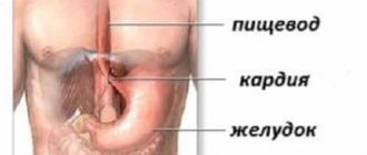
GERB is a chronic disease characterized by periodic exacerbations, during which the stomach contents are released into the patient’s digestive tract. The main reason for such emissions is a weak sphincter - a valve that does not completely block the sections of the gastrointestinal tract from each other. Externally, this is manifested by the replacement of flat and multilayer epithelium with cylindrical epithelium.
Such changes lead to disturbances at the cellular level: the structure of the nucleus and growth are disrupted. Because of this, this pathology becomes the main cause of cancer development. Cylindrical cell metaplasia of the esophagus is one of the main factors in the development of cancer, which is more common in men. If you are overweight, you are much more likely to develop cancer.
What is epithelium?
The gastric epithelium is a collection of columnar cells. They are called single-layer columnar epithelium. Against the background of prolonged irritation of the mucous membrane of the esophagus by gastric juice, for example, with gastroesophageal reflux, when the contents of the stomach are thrown into the esophagus, the risk of developing the gastric variant of esophageal metaplasia increases.
The essence of the disease is the replacement of the stratified squamous epithelium of the esophagus with a single-layer cylindrical epithelium, characteristic of the stomach. The part of the esophagus in which the mucous membrane of the organ passes into the stomach is most susceptible to tissue replacement. It is in the area of connection of the two organs that the most intense irritation with hydrochloric acid occurs against the background of gastroesophageal reflux.

Risk factors
Cylindrical cell metaplasia of the esophagus occurs under the influence of the same provoking factors that lead to the appearance of gastroesophageal reflux disease. Experts identify the following risk factors:
- Decreased tone of the esophageal sphincter in the lower part. This occurs due to insufficient innervation, when the sphincter does not compress tightly and gastric juice is thrown back into the esophagus.
- Changes in the structure of the congenital type. Under the influence of genetic factors, the diameter of the lower part of the esophageal sphincter increases or decreases.
- Previously experienced inflammatory processes. Localized in the lower part of the esophagus, such processes provoke the appearance of scars in the organ and lead to incomplete compression of the sphincter.
- Varicose veins in severe form. In this case, it is the nodes formed during varicose veins that prevent the sphincter from completely closing.
- Disorder of the motor-evacuation function of the stomach and esophagus. Against the background of such a disorder, antiperistalsis is observed, when the walls of the organs begin to move, leading to the movement of food masses into the upper parts of the esophagus.
The listed factors can provoke the appearance of gastroesophageal reflux, which in turn can cause erosive type gastritis, which later develops into gastric and intestinal metaplasia of the esophagus.

Causes
So, the process of gastric metaplasia is a change in the structure of organ tissues. The epithelial cells of the gastric mucosa change, acquiring the properties of small intestinal cells. The replacement process begins against the background of various gastrointestinal diseases, in which epithelial cells die.
Thus, metaplasia is not an independent disease, but a consequence of other pathologies. The mechanism of cell degeneration is as follows:
- the mucous membranes are regularly exposed to unfavorable factors - poor nutrition, exposure to Helicobacter pylori bacteria, uncontrolled use of medications, etc.;
- the response to constant irritation is the development of an inflammatory nature, over time the inflammation becomes chronic;
- Chronic inflammation causes the death of cells and glands, a decrease in the production of digestive juices, and the digestion process is disrupted. In place of atrophied cells, new ones are formed, characteristic of the covering of the small intestine. At this stage, called metaplasia, the pathology is reversible; with proper treatment, the prognosis for recovery is good;
- If left untreated, metaplasia progresses to the next, irreversible stage, called dysplasia. This condition is considered precancerous;
- The last stage is the development of cancer, which often ends in death.
Thus, the reasons for the development of metaplasia are as follows:
- inflammatory chronic processes in the epithelial layer of the stomach;
- inflammatory diseases of the esophagus with subsequent spread to other organs of the digestive system;
- hormonal disorders;
- severe stress;
- bile stagnation, bile reflux.
Advice! Most inflammatory diseases of the stomach are caused by the bacteria Helicobacter pylori. Therefore, during the examination, it is necessary to do an analysis for this infection.
Symptoms
Many people are interested in how the disease manifests itself?
If we are talking about focal metaplasia of the esophagus, then clinical manifestations of this disease, as a rule, are not observed. Suspicion of metaplasia arises from clinical manifestations of gastroesophageal reflux. Therefore, we can say that the signs of the diseases coincide and are as follows:
- Frequent occurrence of heartburn. A strong burning sensation occurs in the retrosternal region, provoked by irritation of the epithelial layer of the esophagus with the contents of the stomach during the reverse reflux.
- Sour belching. Most often it is a precursor to heartburn. Belching appears after eating or while bending the body.
- Pain in the chest area. The intensity of the pain syndrome increases on an empty stomach and tends to radiate to the area of the neck, lower jaw and left half of the chest, as well as to the space between the shoulder blades.
- Extraesophageal signs. This may include shortness of breath, cough, dryness and sore throat, hoarseness, feeling as if the stomach is full even after a small amount of food.
When these signs appear, you should be examined for the presence of foci of metaplasia of the gastric mucosa into the esophagus and determine their extent and severity.
Chronic gastritis with focal metaplasia
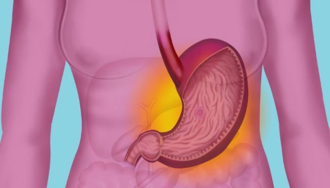
Chronic gastritis is inflammation of the mucous tissues of the stomach, impaired formation of gastric acids with increased or decreased fermentation. Low acidity forms the most dangerous forms of the disease, leading to the death of glandular cells, often leading to a malignant process; if you do not undergo medical examination and treatment on time, it can lead to death.
Chronic gastritis is considered a disease of civilization. Eating heavy, unhealthy foods, abuse of alcohol, carbonated drinks and other harmful substances leads to consequences such as the development of various pathologies of the digestive system.
The working classification of chronic gastritis according to morphological characteristics identifies one of its pathological varieties - gastric metaplasia. Chronic gastritis with focal metaplasia is a pathology in which the cells of the gastric tissue, under the influence of unfavorable causes, are damaged, in place of which cells appear that are similar to another organ: the large or small intestine.
Subsequently, these cells perform their physiologically established function, stopping the function characteristic of the stomach. This disease is rarely diagnosed due to the specific changes in the gastric mucosa.
The period of exacerbation of metaplasia is capable of malignant degeneration.
Characteristic
Metaplasia is more often characterized as a state of transition of the pathological process of the stomach, rather than as a separate disease.
When the human body is exposed to unfavorable factors: alcohol, poor diet, medications and others, gastric tissues can change their appearance. Since tissue restoration usually occurs under the influence of the regenerative process, epithelial cells are renewed faster. The impact of negative factors provokes a failure in cell formation, and the epithelium of the small intestine appears in the stomach. The acute stage of metaplasia provokes replacement with colon cells. This form of the disease is called intestinal metaplasia.
Most often, lesions occur in older people, due to a weakened body, more frequent exposure to various diseases, and exposure to medications.
During the process of replacing gastric cells, two types of pathology are diagnosed:
- Small intestinal metaplasia, incomplete, is not so dangerous; if treatment is carried out in a timely manner, it can be completely cured.
- Colonic metaplasia, complete, often provokes a stomach tumor. With early diagnosis and treatment, this pathological process can be stopped. But you shouldn’t take the disease lightly.
First, an inflammatory process of the mucous membrane develops, which becomes chronic. After cells lose their functional ability, they atrophy. Without timely therapeutic measures, the process moves to the dysplasia stage with the formation of malignancy.
Degeneration of the epithelium can occur throughout the stomach, damage to part of the mucosa or throughout the entire thickness. The location of the lesions is on the body shell, fundus, pyloric, antral sections.
According to statistical observations, doctors have confirmed that the changes caused by focal metaplasia and gastric cancer of the intestinal type are completely identical.
There is a theory that malignancy develops due to carcinogens. It has been recorded that lesions are more common in difficult epidemiological areas.
Causes
The factors causing this process are as diverse as the causes that provoke inflammation itself:
- chronic gastritis caused by Helicobacter bacteria;
- gastroesophageal reflux;
- failure of the pyloric region, reflux of bile from the stomach;
- irritation of mucous membranes;
- hormonal imbalances.
Modern medicine already has evidence of the enormous influence of the Helicobacter bacterium on the development of intestinal metaplasia and malignancy of the digestive tract.
It is this microorganism and its destructive consequences that cause severe tissue degeneration. The Helicobacter bacterium is a class 1 carcinogen.
Types of metaplasia
The disease happens:
- full-small intestinal, mature;
- incomplete - colon, immature.
With mature metaplasia, the presence of cells found only in the small intestine: border cells, sulfamucin, goblet enterocytes. The main feature confirming this variety is Paneth cells. The gastric epithelium resembles the tissue of the small intestine in structure and functionality.
Immature metaplasia provokes impaired maturation and development of the gastric glands - there are almost no differences between the upper and lower layers, the tissue structure consists of cells of the large intestine.
Complete intestinal metaplasia is more common in patients with chronic gastritis and precedes colonic metaplasia.
Metaplasia is divided according to pathological type:
- Pyloric - replacement of the tubular glands of the body of the stomach with mucous tissues - Sterck's pyloric glands (concomitant atrophic gastritis).
- Ciliated - ciliated cells in the stomach, healthy people do not have them, it is believed that their appearance is associated with pathology. Such cells are also found in adenocarcinoma. However, this type of disease does not always cause malignancy.
- Pancreatic - rare, characterized by a fine-grained texture, focal, diffuse form.
Additionally, with pyloric metaplasia – focal, diffuse form of anomalies:
- In focal - some tubular glands are replaced due to inflammation, damage to cell renewal of the gastrointestinal tract.
- Diffuse - characterized by damage to the mucous membranes without structural disturbances or cell necrosis.
Symptoms
To determine the pathology of gastric metaplasia, you need to familiarize yourself with the symptoms and causes of its occurrence.
Gastritis? Ulcer? To prevent a stomach ulcer from turning into cancer, drink a glass...

The best FOLK remedy for GASTRITIS and stomach ulcers!
Transformation of gastric tissue can be triggered by factors:
- various substances, stomach irritants, lowering acidity, as a consequence of exposure - glandular cells of the mucous membranes die;
- irritation of mucous tissues by rough, spicy food, alcohol can cause the development of the disease;
- inflammation of the stomach lining;
- chronic gastritis – often causes the development of metaplasia;
- gastroesophageal reflux;
- neuropsychic overload, stress.
The above reasons for the formation of a pathological process in the stomach, accompanied by a partial or complete stop in the production of gastric juice, acid fermentation necessary for digestion, as a consequence - the death of glandular cells, the formation of metaplasia of mucous tissue instead.
Chronic gastritis with metaplasia in its manifestations is similar to the symptoms of inflammation caused by a decrease in secretory levels. So far no one has been able to describe the symptoms more accurately from medical specialists; there is no clear description of the signs.
Often, due to the similarity of symptoms with other diseases, symptomatic self-treatment is carried out, which cannot be done: external manifestations are weakened, but the disease itself progresses, causing the risk of developing malignancy.
Classic symptoms:
- nausea, vomiting;
- lack of appetite;
- unpleasant sensations in the epigastrium.
Diagnostics
Standard diagnosis using histological examination, small pieces of tissue taken from the human body.
Method of collecting material - biopsy is a mandatory method of diagnosing malignancy.
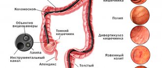
To determine the lesion, an additional examination of the digestive tract is carried out using an endoscope, staining the cells with a special dye that is safe for human health. Damaged tissues become of a special color and are noticeable when viewed under a microscope. Comprehensive diagnostics gives a more accurate picture of the disease. It is also easier to identify the bacteria that cause chronic gastritis with focal metaplasia in order to prevent a precancerous condition.
Therapy
Treatment is selected taking into account damage to the mucosa. With the identified pathology, the patient should be registered with a gastroenterologist.
Drug therapy should initially eliminate:
- gastroesophageal reflux disease - during which the acidic contents of the stomach are regularly thrown back into the esophagus and the acid damages the mucous membrane;
- suppressed gastric secretion;
- neutralization of Helicobacter bacteria;
- benign neoplasms - the treatment plan is selected taking into account the examination results.
Usually prescribed:
- Proton pump inhibitors - reduce the acidity of gastric juice.
- The use of drugs: rabeprozole, omeprozole, pantoprazole, antacids: Maalox, phosphalugel - neutralizes the effect of hydrochloric acid.
- Histamine blockers: cimetidine, ranitidine - antisecretory drugs.
- Gastroprotectors – stops damage to the mucosa when stomach acidity increases
During pregnancy and childhood, there are restrictions on some medications.
- To enhance the effect, it is recommended to take antibacterial drugs: amoxicillin, clarithromycin.
If therapy is not effective, treatment with tetracycline or metronidazole is used. Inhibitors normalize acidity, reduce mucus viscosity, and neutralize the side effects of antibiotics.
- Medicines that boost immunity and prevent dysbacteriosis.
Surgical methods are recommended when conservative treatment is ineffective.
For minimal trauma, a minimally invasive operation is performed using endoscopic equipment, with limited damage to the mucous membrane. If necessary, remove the entire damaged area. The result is a reduction in the risk of carcinogenic formations.
Following a diet is the key to successful treatment.
- It is recommended to exclude from the diet: dairy, fried, spicy, salty foods, alcohol, coffee, carbonated drinks.
- The daily diet should be multiple, small portions.
- Eating fresh vegetables and fruits.
- Cereal porridge.
- It is recommended to eat warm food - hot food irritates the mucous membranes of the stomach, cold food takes a long time to digest and promotes the release of hydrochloric acid.
Patients with metaplasia are registered with a gastroenterologist. Every year they need to undergo FGDS and biopsy for prevention.
Complications
Replacing esophageal tissue with single-layer columnar epithelium can cause a number of serious complications, including:
- Transformation of foci of metaplasia into malignant neoplasms. The most common form of esophageal cancer is adenocarcinoma.
- Hemorrhagic syndrome localized in foci of metaplasia and damage to the esophagus.
- The appearance of peptic-type strictures. These are adhesions of connective tissue that narrow the lumen of the esophagus at their location and provoke the development of dysphagia, which consists in disrupting the process of swallowing food.

To prevent such complications, it is important to diagnose metaplasia in time and receive appropriate treatment aimed at reducing the size of the lesions and their number.
Diagnostics
The diagnosis of columnar cell metaplasia of the esophagus is made only on the basis of an instrumental examination of the patient. Modern medicine uses several methods to visualize the mucous membrane of the esophagus, including:
- Esophagoscopy. An instrument called an endoscope, which is a fiber optic tube equipped with lighting and a camera, is inserted into the lumen of the organ. The doctor can assess the condition of the esophageal mucosa by receiving an image from an endoscope on a special monitor. To obtain a more detailed image of metaplasia, a contrast agent is used to highlight foci of the pathological process. In this way, it is possible to establish the presence of an oncological neoplasm and identify metaplasia at the initial stage of development.
- Targeted biopsy. It is carried out along with the previous diagnostic method. Esophagoscopy determines the exact location of the foci of metaplasia, which allows tissue to be taken for histological examination specifically from the affected area. A specialist examines the resulting sample in a laboratory setting, determining the presence of single-layer columnar epithelium in the esophageal tissue.
- X-ray examination. Helps to visualize the structures of the walls of the esophagus and assess its motor-evacuation ability. This method also uses a contrast agent injected into the lumen of the esophagus.
- Manometry. It is carried out by measuring pressure in the esophageal lumen.
- Impedancemetry. Study of the motor-evacuation ability of all parts of the esophagus.
After a thorough examination and clarification of the diagnosis, the doctor prescribes treatment for metaplasia.

Treatment: general principles
The basic principles of treatment of esophageal metaplasia are the elimination and reduction of the size of foci of a single-layer columnar epithelial layer in the esophageal mucosa. Therapy is carried out comprehensively and includes both conservative treatment and surgical methods, and general recommendations on nutrition and lifestyle.
Experts believe that an important stage in the treatment of gastric metaplasia of the esophagus is the implementation of general recommendations that allow you to relieve the symptoms of the pathology. In addition, following the rules listed below increases the effectiveness of other treatment methods:
- Maintaining a balanced diet. Hot and cold dishes, as well as fried, fatty, smoked foods, marinades, pickles and spices are excluded.
- Supporting proper nutrition. You should eat at least five times a day in small portions. The final meal should be at least two hours before going to bed. You should try not to lie down after eating.
- Bringing body weight back to normal, subject to its deficiency or excess.
- Quitting bad habits, including alcohol and smoking. Such habits contribute to even greater irritation of the mucous membrane of the esophagus and increase the risk of increasing lesions of the disease.
- Physical activity should be moderate. Lifting heavy objects should be avoided as this increases the risk of developing pressure within the peritoneum.
The listed rules will increase the effectiveness of the treatment and prevent surgical intervention by eliminating small foci of gastric metaplasia of the esophageal mucosa.

Can Barrett's Esophagus be cured? (Issue 4)
Barrett's esophagus is defined as the replacement of the esophageal mucosa by gastric mucosa of columnar cell metaplasia with the presence of foci of intestinal cells of intestinal metaplasia among it. If the development of columnar cell metaplasia has already taken place, is it possible to ensure that normal esophageal epithelium forms in this place during treatment? Many studies have been devoted to solving the question of whether Barrett's esophagus can be cured and, unfortunately, today neither drug nor surgical treatment can provide this with evidence-based reliability.
A number of studies have noted the appearance of even islands of new esophageal epithelium, but not its complete restoration. Some authors reported complete regression of columnar cell metaplasia, but only in 2 patients observed. Is it possible to cure Barrett's esophagus by destroying the foci of the disease? Various forms of ablation, destruction of foci of Barrett's esophagus by various methods, are currently considered a promising and modern method of treating Barrett's esophagus.
These methods are resorted to only when Barrett's esophagus progresses, that is, when it is detected against the background of columnar cell metaplasia, intestinal metaplasia in combination with dysplasia.
It should also be noted that these procedures are done under the control of human vision, not at the microscopic level. Not isolated cases of residual microscopic foci of Barrett's esophagus that were not destroyed have been described. A new epithelium of the esophagus was formed on top of these foci, and underneath it, from the undestroyed remnants of Barrett's esophagus, a process continued to develop, which was not determined in any way during subsequent endoscopy.
Can Barrett's Esophagus Be Cured by Changing Your Mindset? Therefore, answering this question, today we can say with confidence: medicine is not yet capable of this. If Barrett's esophagus is suspected and detected, it is necessary to carry out lifelong drug therapy with an optimal combination of surgical interventions, and if dysplastic processes occur in the mucous membrane of the Barrett's esophagus, resort to destruction, i.e.
Gastric metaplasia or precancerous condition.
Drug treatment
Conservative treatment of esophageal metaplasia involves taking medications whose action is aimed at reducing the amount of gastric juice reflux into the esophagus. For this purpose, specialists prescribe the following groups of medications to patients:
- Antacids. The effect of these drugs is to reduce the acidic qualities of gastric juice. Antacids include Maalox, Almagel, Phosphalugel, etc.
- Proton pump blockers. These drugs inhibit the production of hydrochloric acid. The most popular drug in this group for esophageal metaplasia is Omeprazole.
- H1-histamine receptor blockers. Also used to reduce the amount of acid produced. The most commonly prescribed drug is Famotidine.
- Prokinetics. These drugs have a stimulating effect on the peristalsis of the stomach and esophagus, which prevents the reflux of food mass into the upper part of the digestive organs. One of the brightest representatives of this group of medicines is Motilium.
The blockers mentioned above are prescribed only after checking the acidity of the gastric juice, as well as determining the intensity of its peristalsis. Only small foci of metaplasia of the esophageal epithelium can be treated with medication.
Diet
Treatment will not be complete without proper nutrition. Patients are recommended:
- exclude dairy products from the menu, as well as those that irritate the gastric mucosa (fried, spicy, salty);
- do not drink alcohol, coffee, carbonated drinks;
- meals should be fractional, at least 5–6 rubles/day;
- last meal – 2–3 hours before bedtime;
- The diet must include fresh vegetables and fruits.
Nutritionists advise introducing a variety of cereals into your diet. It is recommended to eat food only warm.
Too hot food irritates the gastric mucosa, while cold food takes a long time to digest and provokes the release of hydrochloric acid.
Photo gallery: foods you can eat
Surgery
This is a radical treatment option and is considered a last resort. Surgeons use several techniques to remove esophageal metaplasia, including:
- Open access surgery. This is a traumatic manipulation when the chest is dissected and the affected part of the esophagus is removed. The operation is prescribed in cases of extensive damage to the esophagus by foci of metaplasia.
- Fundoplication. This operation is also performed with an open approach, but during fundoplication the gastric fundus is sutured to the diaphragm. In this way, it is possible to form an acute angle with the esophagus and reduce the reflux of stomach juice into the organ.
- Laser therapy. This is a more modern method than abdominal surgery. Using a low-frequency laser, the single-layer cylindrical epithelial layer in the area of metaplasia formation is destroyed.
- Photodynamic therapy. Foci of metaplasia are destroyed by introducing a special photochemical element into them. Next, the substance is exposed to light of a certain wavelength, which leads to the death of unnecessary cells.