Can atrophy of the gastric mucosa be treated, how can it be treated and restored so that the symptoms of the disease do not reappear? Gastric atrophy is a process of pathological origin that occurs after inflammation. Healthy stomach cells are replaced by connective tissue, which becomes thinner over time. Issues related to diagnosis and treatment are discussed in detail in this article.

Classification of the disease
Today there are several classifications of mucosal atrophy, which are based on the stage of development of the pathology and the severity of its symptoms.
Classification according to the degree of development of pathology:
- subatrophic: the initial stage of pathology development;
- focal: the first areas appear in which the number of glands is significantly reduced;
- atrophic antral gastritis: a progressive stage of the disease in which the lesion occurs in the antrum of the stomach;
- multifactorial: pathology spreads throughout the entire mucous membrane of the organ.
Classification of atrophic gastritis according to the degree of symptoms:
- moderate;
- expressed;
- hyperplastic;
- with the appearance of fatty degeneration and cysts.
Distal reflux esophagitis
Distal reflux esophagitis occurs against the background of permanent irritation of the esophagus. The tissues of the organ located closer to the stomach gradually begin to become inflamed, hyperemia of the mucous membrane and swelling appear. A feature of the disease is the duration of its course; the initial stages rarely manifest any symptoms. As a result, distal esophagitis of a chronic nature develops, which is difficult to treat and often recurs.
The main source of distal inflammation of the esophagus is insufficiency of the cardia, that is, the circular muscle that blocks the entrance to the stomach. When the sphincter does not close tightly, the acidic contents of the stomach are transferred to the lower esophagus, causing irritation of the mucous membrane and severe inflammation. Often, the cause of distal GERD is gastrointestinal diseases, which are accompanied by increased stomach acidity, such as gastritis.
Distal esophagitis is characterized by inflammation of the esophageal mucosa of varying severity. Very often, distal reflux esophagitis appears during repeated cycles of cardia deficiency and occurs against the background of the influence of acidic gastric contents on the esophagus. Diagnostic measures for distal esophagitis require analysis of symptoms and the results of a complete examination of the gastrointestinal tract.
The main source of distal inflammation of the esophagus is insufficiency of the cardia, that is, the circular muscle that blocks the entrance to the stomach
Drug therapy for distal type esophagitis includes antifungal and enveloping drugs, antacids and antispasmodics. In addition, peloid therapy (mud treatment) and electrotherapy (amplipulse therapy) have proven themselves. Reflux indicates degenerative changes in the body.
Gastritis distal catarrhal
Distal catarrhal gastritis is common among men. Inflammation reaches the far walls of the stomach due to consumption of spicy foods or alcohol damage. The distal form is chronic and erosive. The presence of a feeling of heaviness, attacks of nausea and bloating after eating warns a person that inflammation requires immediate investigation and treatment.
Focal distal gastritis and catarrhal superficial distal gastritis
A form of the disease in which individual foci are affected - islands of the mucous membrane. This occurs due to local damage to the shell structure either by the bacterium Helicobacter pylori or poisoning. In the affected areas, cells responsible for the production of digestive enzymes die. This leads to headaches, stomach pain and vomiting. Catarrhal superficial gastritis, in which inflammation affects exclusively the upper layers of the epithelium, without causing harm to the deeper ones.
Pathogenesis
Before talking about the diagnosis and treatment of this disease, a few words should be said about its pathogenesis and describe the processes that lead to degeneration of the mucous membrane of the organ.
Today it is believed that two factors lead to the development of this pathology:
- exposure to microorganisms;
- autoimmune processes.
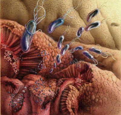
For convenience of description, we divide the pathological process into two stages. At the initial stage, acid-fast bacteria, which are common inhabitants of our stomach, play a key role. First of all, we are talking about Helicobacter pylori, a bacterium that is the main cause of the development of most forms of gastritis.
These microorganisms damage the protection of the cells of the mucous layer and change the pH of the environment around them. We can say that a bacterial infection only prepares the ground for the further development of pathology, which can take different paths, including atrophy.
At the second stage of pathogenesis, autoimmune processes come into play, which do not allow new cells of the mucous membrane to differentiate. New cells, instead of gastric juice, produce mucus, which does not participate in the digestive processes.
The walls of our stomach have a high ability to regenerate; the cells of the mucous membrane are renewed every six days.
The secretion of hydrochloric acid is provided by parietal cells. The body begins to produce antibodies against them, which bind to the microvilli of the cells. They interfere with the absorption of vitamin B12 and disrupt the functioning of the H+/K+-ATPase ion pump, the main mechanism that synthesizes hydrochloric acid.
Severe gastric atrophy of the antrum
Hydrochloric acid is directly involved in the digestive processes that occur in the stomach. It denatures food proteins and activates the digestive enzymes of this organ. Hydrochloric acid also performs a bactericidal function in the stomach.
The transformation of atypical cells of the mucous membrane into malignant ones does not always occur; this process depends on a large number of factors. Our body has several defense mechanisms; the likelihood of onset of cancer depends on the effectiveness of their functioning.
Summarizing the above, we can conclude that atrophic gastritis is the result of long-term Helicobacter pylori gastritis and a special case of autoimmune gastritis.
In some cases, mucosal atrophy is entirely due to autoimmune mechanisms, but this is observed extremely rarely, mainly in young patients.
Prevention and prognosis of atrophy of the gastric mucosa
Focal superficial gastritis is a lesion of the gastric mucosa, in which only one of the sections of the stomach is involved in the process - the fundic or pyloric. This is the initial, mildest stage of the disease.
The most common forms of superficial focal gastritis are antral and distal (pyloric) localization.
The main cause of the disease is Helicobacter pylori. Its symptoms are mild or not expressed at all. The main complaint of patients is epigastric pain.
Frequent symptoms of the disease are: lack of appetite, heartburn, belching, flatulence, stool disorders. Treatment of superficial gastritis includes diet, medications (usually in the form of tablets), herbal medicine, and normalization of lifestyle. With timely treatment, the prognosis is quite favorable.
But patients often do not seek help, and the disease progresses. Cellular elements are activated in the coolant, causing secondary damage to epithelial cells - gastric epithelial cells. Chronic erosions occur, often with a hemorrhagic component.
How to treat erosive gastritis and what medications are used
Further activation of the bacterium Helicobacter pylori provokes the development of mucosal atrophy, and the processes of cellular regeneration are disrupted. A mixed form of gastritis occurs, represented by both superficial and atrophic forms. As a result, bicarbonates, necessary for alkalizing food before entering the intestines, cease to be produced.
Symptoms of duodenitis and increased pressure in the duodenum occur. The closing function of the pylorus suffers, and DGR occurs. The effect on the antral mucosa of refluxate (which includes bile acids, salts of pancreatic enzymes, duodenal contents) causes reflux gastritis.
Symptoms
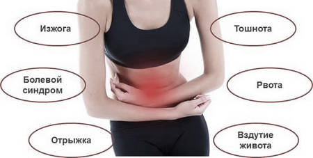
Disruption of the normal functioning of the gastric glands has an extremely negative impact on the processes of food digestion that occur in the stomach. A characteristic symptom of atrophic gastritis is dyspepsia, in this it differs from the hyperacid variety of this disease, where the main manifestation is pain symptoms.
Typical symptoms of dyspepsia include:
- nausea;
- loss of appetite;
- frequent belching;
- smell from the mouth;
- heaviness in the stomach;
- bloating;
- diarrhea;
- salivation.
Atrophic gastritis may be accompanied by pain, but for this disease they are less characteristic than for other types of gastritis.
A decrease in the amount of hydrochloric acid leads to disruption of the organ. Food stays in the stomach longer, is less digestible, and a decrease in acidity leads to an increase in the number of bacteria (hence intolerance to dairy products and diarrhea). In addition, disruption of the normal functioning of the stomach negatively affects the functioning of the entire digestive tract and can contribute to the development of diseases of other organs: pancreatitis, peptic ulcer, cholecystitis.
Atrophic gastritis is accompanied by other symptoms not related to the functioning of the digestive system:
- anemia;
- weight loss;
- hormonal imbalance (decreased libido).
Anemia develops due to impaired absorption of vitamin B12, which can also lead to mouth ulcers, yellowing of the skin and frequent headaches.
Weight loss, often seen with this disease, is a consequence of diarrhea and the insufficient amount of nutrients (mainly proteins) that the body receives with this disease. Painful sensations, which are usually aching in nature, arise due to a violation of the evacuation function of the stomach and its stretching.
The initial stages of all types of disease are almost always asymptomatic.
Superficial gastritis: symptoms in adults
Superficial gastritis can occur for a long time without visible signs - asymptomatic.
The main symptoms of superficial gastritis in adults:
- Discomfort in the epigastrium after eating difficult to digest or poor quality food. Some patients complain of dull pain that is diffuse in nature.
- Heartburn.
- Nausea, belching.
- Frequent constipation.
Women often face the problem of gastritis during pregnancy. The disease itself, although it does not affect the development of the fetus, causes discomfort in the woman, and adequate treatment during this period is very difficult.
Symptoms of superficial gastritis in adults
Antral gastritis in adults is most often associated with Helicobacter pylori and causes gastroduodenitis (combined inflammation of the stomach and duodenum). This infection provokes the transition of mild forms of the disease to atrophic gastritis, which threatens metaplastic degeneration of the mucosa and even cancer.
Chronic superficial gastritis associated with Helicobacter pylori
Causes of pathology
The main cause of the disease is Helicobacter pylori infection. Usually, mucosal atrophy is a consequence of an infectious form of gastritis that has lasted for a long time.
Hereditary factors play a major role in the development of this pathology. The likelihood of this disease occurring is significantly increased by non-compliance with the diet and too frequent consumption of foods that irritate the gastric mucosa. Drinking alcohol and smoking have a very negative impact on the condition of the gastric mucosa. Various previous infections and stress also increase the likelihood of developing the disease.
Consequences of the disease
Atrophy of glandular cells leads to disruption of the normal functioning of the organ. This disrupts the functioning of the digestive system as a whole. However, the greatest danger posed by this pathology is the occurrence of gastric cancer.
Gastric atrophy is considered a precancerous disease; this disease is often a precursor to carcinomas. The most dangerous in this regard is chronic atrophic gastritis, with which the likelihood of developing cancer is 15%.
In addition to the development of neoplasms, atrophy of the mucosa is usually accompanied by disturbances in the functioning of other organs of the digestive system: the duodenum is especially affected.
Animal organ[edit | edit wiki text]
Section source:
Biological encyclopedic dictionary
[1]
In invertebrates[edit | edit wiki text]
The stomach as a separate section is already present in many invertebrates. In many arthropods, food is ground in the stomach. Among crustaceans, the lower ones have a glandular stomach, while the higher ones have a chewing stomach. In arachnids, the stomach is represented by the first section of the midgut with blind appendages. Insects have a well-developed chewing stomach; only some forms have a glandular stomach. In mollusks, digestive glands (“liver”) open in the stomach. Among echinoderms, the stomach is developed in crinoids, starfish and brittle stars. A separate stomach is present in some hemichordates and tunicates.
In chordates[edit | edit wiki text]
In general, the evolution of the stomach, associated with the ecological specialization of species, followed the path of increasing complexity of the shape and structure of the stomach membranes that make up its wall (mucous, muscular and serous).
In cephalochordates and fish[edit | edit wiki text]
Among typical chordates, the lancelet, cyclostomes and some fish do not have a separate stomach. In shark fish, the stomach is curved in a horseshoe shape; there is a cardiac part, which extends from the esophagus, and a pyloric part, which passes into the intestine; Between them is the fundus of the stomach. In bony fish, blind pyloric outgrowths extend from the intestine near the stomach. More often, the stomach of fish is not clearly defined.
In amphibians and reptiles[edit | edit wiki text]
The stomach is more clearly separated in amphibians and reptiles.
In birds[edit | edit wiki text]
In birds, the stomach is divided into 2 sections: the muscular section, in which the muscular layer is highly developed, and the mucous membrane is covered with stratified squamous keratinizing epithelium, and the glandular section, the mucous membrane of which is equipped with branching glands. The powerful muscles and cuticle of the muscular stomach, together with swallowed small pebbles and grains of sand (the so-called gastroliths), contribute to the mechanical processing of food, compensating for the lack of teeth in birds.
In mammals[edit | edit wiki text]
The stomach reaches significant development in mammals. Their simple single-chamber stomach has 4 sections: esophageal, adjacent to the entrance of the esophagus, cardiac, bottom and pyloric, lined with glandular epithelium, forming the corresponding glands. In different mammals, due to food specialization, the degree of development of the departments is not the same. Thus, in monotremes, the single-chamber stomach is entirely lined with multilayered epithelium - it is glandless, of the esophageal type. In cetaceans, the areas of the benthic and pyloric glands have grown and separated, forming a multi-chambered stomach. In omnivores, the esophageal region is well defined, and the area of the cardiac glands has grown and separated in the form of a diverticulum. In ruminants, the esophageal section has reached its greatest development, from which three chambers of the proventriculus arose: the booklet, the mesh, the rumen, and the fourth section, the abomasum, is the glandular stomach itself. Carnivores and humans have a single-chamber stomach of the glandular type with minimal esophageal lining and well-developed cardiac, fundic and pyloric glands. The capacity of the human stomach is on average 1.5-2.5 liters; in men it is slightly larger than in women.
Gallery[edit | edit wiki text]
Digestive organs - main functions
- Topography of the abdominal organs
- Topography of the abdominal organs. Sagittal section. Left view
- Anatomy (structure) of the stomach
- Structure of the stomach
- Cross section of the stomach wall
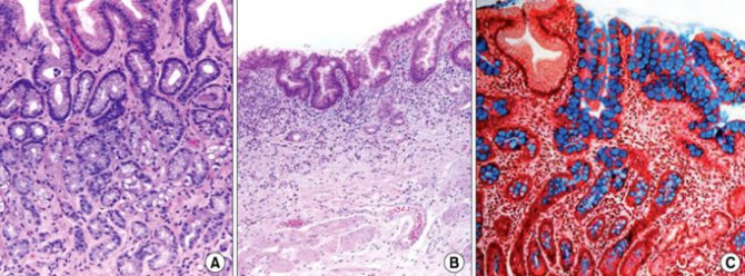
Cells of the fundus of the stomach, structure of the gastric glands, cells of the glands of the fundus of the stomach
Diagnostics
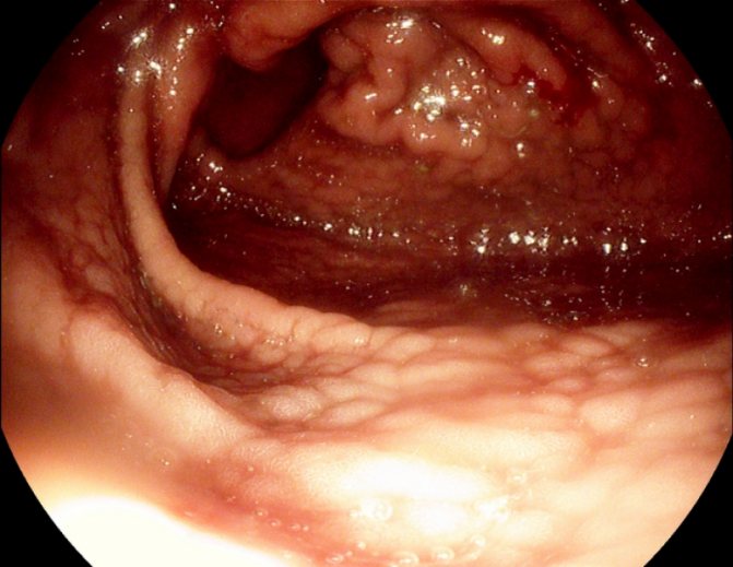
Diagnosis of this pathology is quite difficult. It is especially difficult to distinguish this disease from stomach cancer.
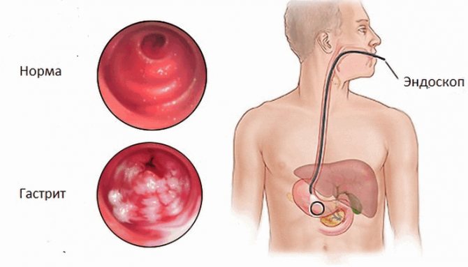
Mucosal atrophy visible on endoscopy
Ultrasound, X-ray methods, computed tomography and MRI cannot give a definitive answer regarding mucosal atrophy. Endoscopic methods are more effective; chromogastroscopy is sometimes used.
Initially, basic tests are taken from the patient. The blood test is especially interesting: with a lack of vitamin B12, a decrease in hemoglobin levels is often observed.
A mucosal biopsy should be performed, which can show the type of tissue changes. It is also necessary to study the pH value of gastric juice; with atrophic gastritis it is usually lowered.
In addition, antibodies to Helicobacter pylori are detected, and the level of pepsinogen and gastrin proteins, which are involved in the synthesis of hydrochloric acid, is studied.
Structure of the stomach
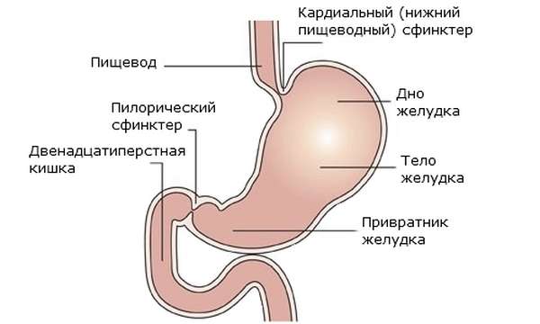
The stomach is a hollow organ that is located in the upper part of the abdominal cavity. It begins at the place where the lower edge of the esophagus passes into the cardiac part of the stomach (approximately at the level of the 10th thoracic spine). There is a sphincter here that prevents food from being thrown back into the upper digestive tract.
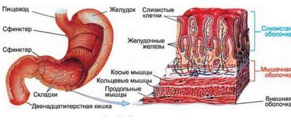
The cardiac section expands and passes into the body - the main part of the organ. This is where the main processes of digestion and grinding take place. The bottom extends slightly upward from the body - an area where air often accumulates. Below, the body gradually begins to narrow and passes into the pyloric region. Between it and the duodenum there is the pylorus - a powerful smooth muscle sphincter that regulates the passage of food masses.
The wall consists of several layers:
- The mucous membrane is formed by columnar epithelium. Below it is the lamina propria, which contains connective tissue and glandular cells.
- Smooth muscle - consists of three balls of elastic muscles, which are located transversely to each other. This provides greater extensibility of the walls of the organ. Regular peristaltic movements greatly crush the food mass.
- The adventitia , which is almost completely covered by the peritoneum.
The normal shape of the stomach is horn-like. They also distinguish between the greater and lesser curvature, the anterior and posterior walls of the organ.
Treatment
Treatment of the pathology depends on the reasons that caused it, its type and stage of the disease. If it is based on an infection caused by Helicobacter pylori, then eradication therapy is used. Its main task is to destroy pathogenic microflora.
Before starting medication, a daily measurement of the pH level of the stomach is carried out. If it is less than six, then antibiotics and ion pump inhibitors are used; if the pH value is above six, only antibiotics are used. Amoxicillin and clarithromycin are commonly used.
In some cases, drugs are prescribed that stimulate the production of hydrochloric acid, as well as drugs that improve organ motility.
The autoimmune mechanisms of the disease are currently not treated.
With this disease, you should not self-medicate, because the likelihood of atrophic gastritis turning into an ulcer or cancer is too high.
Content
- 1 Animal organ 1.1 In invertebrates
- 1.2 In chordates 1.2.1 In cephalochordates and fish
- 1.2.2 In amphibians and reptiles
- 1.2.3 In birds
- 1.2.4 In mammals
- 2.1 Functions 2.1.1 Control over “reception”
- 2.2.1 Psyche