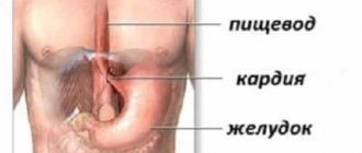
Infection of the esophageal tube with a yeast-like fungus from the genus Candida most often occurs endogenously along the ascending path from the intestines or descending from the oral cavity (more about the symptoms of the disease).
The disease does not occur in isolation; in a significant proportion of cases it demonstrates one of the forms of general candidiasis of all digestive organs.
Therefore, treatment of esophageal candidiasis cannot be carried out in isolation, without identifying and treating other digestive organs. If this condition is not met, candida fungi will constantly remain in the hollow tube, since there is always a risk of infection from other organs. In such conditions, treatment becomes difficult, and it is almost impossible to obtain a positive effect.
Reasons for development
Medical studies show that even completely healthy people are carriers of Candida fungi. It has been revealed that 80% of people have such fungi in the intestines, and more than 25% in the oral cavity. When conditions arise that are conducive to the development of these fungal formations, they begin to multiply vigorously and colonies infect various systems of the human body. Thus, candidiasis affects the esophagus both ascending from the intestine (in most cases), and descending - transferred from the oral cavity.
The population size of this yeast-like fungus rapidly increases in the esophagus and leads to the development of candidiasis. Very often the disease affects other organs of the digestive tract. Since candida fungi are very common in nature, esophageal candidiasis can also begin due to infection from the external environment.
It can begin to develop when:
- contact with the patient;
- use of household or hygiene items;
- eating contaminated food;
- etc.
In order for a disease to develop, it needs favorable conditions. One of the main factors contributing to the development of candidiasis in the esophagus is a weakened human immune system. This type of damage to the esophagus is typical for people with a weak immune system; small children and patients with immunodeficiency often suffer from candidiasis.
Ways of infection with gastrointestinal candidiasis
Fungi of the genus Candida are permanent inhabitants of the human body and are classified as opportunistic microorganisms. Primary infection occurs at the stage of intrauterine development, then the spores of the fungus enter through the esophagus with mother's milk, through surrounding objects, and by airborne droplets from a sick person. It does not matter at what age the infection occurred. The presence of thrush in the microflora does not manifest itself in any way until the immune defense is weakened. The active development of a parasitic fungus causes visceral lesions.
Factors that contribute to the disease
Various forms of disorders also create a favorable environment for the development of the disease:
- anatomical (injury or damage to the esophagus with sharp objects or bones from food);
- physiological factors;
- immunological protective mechanisms.
Factors that provoke manifestations of esophageal candidiasis are:
- use of injected or inhaled corticosteroids;
- long-term use of antibiotics;
- consequences of antacid therapy;
- hypochlorhylric conditions;
- diabetes;
- consequences of intoxication;
- alcoholism and smoking;
- esophageal motility disorder;
- esophageal obstruction;
- malnutrition;
- enteral and especially parenteral nutrition;
- organ or bone marrow transplantation.
Hypofunction of the adrenal glands or parathyroid glands, which lead to disturbances in calcium-phosphorus metabolism, also contributes to the development of candidiasis in the esophagus. Such dysfunction causes a latent form of spasmophilia of the esophagus, which leads to a decrease in its protective capabilities. The prerequisites for the development of candidal lesions of the esophagus are created by a lack of protein, leading to nutritional status. This deficiency can be caused by eating low-calorie foods for too long and all kinds of restrictive diets.
The list of risk factors also includes diabetes mellitus, which leads to an increase in glucose levels in the circulatory system; such hyperglycemia weakens the function of granulocytes.
A decrease in the degree of acidity in gastric juice also contributes to the development of candidiasis. A pH shift to 4.5 completely inhibits the growth of fungi; the optimal environment for their development is a pH level of 7.4.
Check out this article: Candidiasis - burning and pain when urinating
Characteristic symptoms of the disease
In the field of gastroenterology, esophageal candidiasis is one of the most difficult types of disease to identify. For esophageal candidiasis, a very characteristic discrepancy is the severity of the disease, the level of damage to the walls of the esophagus and the sensations of the patient himself. Almost 30% of patients show virtually no symptoms, and even the patients themselves may not even realize that they have such a disease. This is especially true for people who have a reduced level of immunity.
But, nevertheless, the remaining 70% of patients had the following forms of manifestation of this disease:
- heartburn;
- decreased appetite;
- disturbances in the process of swallowing food (dysphagia);
- pain during swallowing (odynophagia);
- chest pain;
- quite frequent vomiting and nausea;
- temperature increase;
- loose stools;
- upper abdominal pain.
Pain when swallowing can be either minor or severe, causing even the inability to eat food and even drink water. This severe condition can lead to dehydration. With symptoms of heartburn, nausea and vomiting, very often characteristic whitish films are observed in the vomit.
Consequences
Late stages of candidiasis can cause erosive, catarrhal gastritis. Symptoms of this condition include pain in the upper stomach and vomiting of blood and white mucus. Candidiasis in the stomach can cause perforation of the stomach wall, which is accompanied by bleeding and the development of peritonitis. If a large blood vessel is damaged, blood loss can be significant.
Bleeding contributes to the spread of fungal spores throughout the body, additional foci of infection appear, and anemia develops. In the later stages of the disease, the mucous membranes are affected very deeply, so conventional drug treatment is not effective. Therapeutic measures involve taking stronger drugs or surgery.
Damaged tissues are not able to withstand pathogenic effects. Gastric candidiasis provokes systemic skin lesions; as the disease worsens, invasive infections and gastrointestinal diseases caused by bacteria occur. The combined effect of all these factors against the background of weakened immune defense can lead to death.
Other signs
Since candidiasis of the esophagus is one of the manifestations of candidiasis that occurs in the gastrointestinal tract, patients often experience loose stools interspersed with mucus and bloody discharge. In such patients, appetite disappears and body weight decreases. Very often, such candidiasis can also manifest itself in lesions of the oral cavity (thrush).
In the initial phase of the development of the disease, the infection penetrates only the surface of the mucous membrane, and then takes it deeper, penetrating into the structure. In this case, characteristic films are formed on the surface. Sometimes they can completely block the lumen in the esophagus. With this disease, necrotic areas, sometimes phlegmons or ulcers, form on the walls of the esophagus, resulting from the addition of infections of bacterial origin to them.
Complications with esophageal candidiasis can occur quite often, but less than with intestinal candidiasis. However, patients experience perforations, development of ulcers, bleeding from blood vessels and tissue necrosis.
What is gastric candidiasis
When the intestinal microflora is disrupted, the parasitic fungus begins to become active. Its spores spread into the oral cavity, esophagus, and stomach. In the normal state of the mucous membranes of the digestive organs, the occurrence of candidiasis is impossible. The internal microflora secretes antifungal components. In the presence of chronic gastritis, stomach ulcers and other inflammations, fungal spores enter the affected areas of the mucous membrane and gradually spread to healthy ones.
Manifestations
Manifestations of esophageal candidiasis come in various forms. At first, the affected areas on the walls of the esophagus look like yellowish or whitish spots that are raised above the mucous surface. Afterwards, such formations can merge and form plaques with the introduction of fungal colonies into submucosal surfaces or pseudomembranous deposits. Fungi penetrate into blood vessels and muscle membranes. Plaque films consist of desquamated epithelial cells that are mixed with fungal bodies, inflammatory cells and other bacteria. Microscopic examinations reveal the characteristic threads of Candida mycelium, which have a yeast-like structure and uniform color.
Check out this article: Traditional medicine successfully fights thrush
The morphological classification of esophageal candidiasis allows us to distinguish the severity of the development of the process, which depends on the depth of damage to its walls:
- group. There are individual whitish plaques with the manifestation of the introduction of pseudomycelium of fungi between epithelial cells;
- group. Film deposits merge with each other and create vast fields. Threads of pseudomycelium penetrate not only into the mucous membrane, but also into the submucosal tissue;
- group. Pseudomembranous deposits are formed, which are combined with a deep phase of changes in which threads of the fungus penetrate into the thickness of the muscle tissue.
Diagnostics
Patients who complain of difficulty swallowing food or associated pain must be examined for the presence of esophageal candidiasis. For this purpose, endoscopic examinations can be carried out using specialized optical equipment, which allows a detailed examination of the mucous membrane of the esophagus (esophagoscopy).
Endoscopic signs of candidiasis in the esophagus are contact vulnerability of the surface of the mucous membrane, hyperemia, as well as the development of fibrous plaques of various configurations, locations and sizes.
There are three groups of manifestations of esophageal candidiasis:
- Catarrhal endophagitis. It reveals moderate swelling of the mucous membranes and diffuse hyperemia (from mild to pronounced). A characteristic symptom is bleeding upon contact with the mucous membrane, as well as the formation of a cobweb-like whitish coating on its surface. Erosion does not appear.
- Psephdomembrane (fibrinous) esophalitis. Loose deposits in the form of rounded plaques up to 5 mm are noted. Noticeably expressed contact vulnerability and hyperemia.
- Erosive fibrous esophalitis. Plaques stand out in the form of fringed ribbons of a dirty gray color, which are located in the esophagus on the crest of the longitudinal folds. Erosion of linear and rounded shapes, up to 4 mm in diameter. The extremely vulnerable mucous membrane has pronounced congestion and swelling. Changes caused by esophageal candidiasis may even interfere with full examination with an endoscope and cause pain, bleeding, or stenosis of the esophagus, which is caused by swelling.
For more severe cases, X-ray examinations using a contrast agent may be used.
For quick diagnosis without the use of an endoscope, special research techniques are used in which instruments are inserted through a protective catheter through the mouth or nose. In this case, traces of mucous remain on the instrument, which are examined in a cytology laboratory. It is also possible to conduct a microbiological study of mucus taken from the esophagus and inoculate it on nutrient media. This analysis allows us to identify the sensitivity of infectious agents to various types of antifungal drugs. For sick people, a study of the state of the immune system is carried out.
Classification of inflammation
A special classification according to the degree of damage to the esophagus facilitates the diagnosis and choice of treatment:
- Catarrhal form: along with a white coating, there is redness, inflammation of the mucous membrane and minimal pain when swallowing.
- Fibrinous: bleeding erosions form under the plaque, swelling causes anxiety and interferes with eating.
- Erosive: the entire mucous membrane is covered with a dense layer of cheesy plaque, large erosions bleed and cause severe redness. Areas of necrosis may be detected.
It is rarely possible to detect the disease at the first stage and almost always by accident.
Treatment
Treatment of esophageal candidiasis is carried out using antifungal (antimycotic) agents and immunostimulants. Antimycotic substances are prescribed based on the results of laboratory tests and identified resistant and non-resistant types of sensitivity to various drugs.
Check out this article: Complications of Oral Candidiasis
Immunostimulating substances are prescribed only after violations in the functioning of the immune system have been identified, since different types of these drugs have different degrees of impact on the functional components of human immunity.
When determining esophageal candidiasis in a person, it is necessary to identify and treat candidiasis in other organs of the digestive system. Without such therapy, it will be almost impossible to treat candidiasis in the esophagus, since there will be constant infection from other organs.
The arsenal of antifungal agents is quite extensive. To treat candidal lesions of the esophagus, oral therapy is initially carried out; intravenous administration of drugs is used in refractory cases of the disease.
The following will increase the likelihood of curing esophageal candidiasis:
- operational diagnosis;
- selection of effective means of specific antifungal therapy;
- carrying out a set of therapeutic measures to stimulate phagocytosis and increase the number of granulocytes.
In the treatment of candidiasis in the esophagus, good results have been obtained through the endoscopic administration of granulocyte concentrates, as well as the use of high-intensity pulses of laser radiation, which improve the immune functions of the human body.
Prevention
Thrush is easier to prevent than to treat. Activation of Candida occurs as a result of weakening of the immune defense, so preventive measures are aimed at improving overall health:
- take antibiotics and drugs that weaken the microflora strictly as prescribed by the doctor;
- follow the principles of healthy eating;
- practice preventive courses of vitamin complexes;
- normalize feasible physical activity;
- promptly treat inflammation and infections;
- do not self-medicate;
- regularly carry out prevention and treatment of chronic ulcers, gastritis, erosion to avoid uncontrolled growth of bacteria and fungi;
- In case of acute manifestations of pathology, it is necessary to undergo a course of antifungal therapy.
Medicinal treatments
For patients with moderate severity of the disease and minimal immune impairment, a shortened course of therapeutic agents is indicated, using absorbable drugs in the form of an oral azole. For the treatment of candidiasis in the esophagus, these substances are most effective. Non-absorbable azoles such as miconazole or clotrimazole are prescribed orally. Substances of this group of systemic action (fluconazole, ketoconazole or itraconazole) have a great effect. These drugs change the degree of permeability of the fungal cell membrane and lead to damage to the cell itself and its death.
Ketoconazole (oronozal, nizoral) is an imidazole derivative; taking 200 mg per day gives a good effect for the treatment of the esophageal canal. The substance is well absorbed in the gastrointestinal tract itself, but for this it needs an optimal acidic environment.
Fluconazole (diflazone, forcan, diflucan and the domestic analogue - flucostat) is a water-soluble form of triazole. It is prescribed 100 mg per day. The degree of absorption of this substance does not depend on the level of acidity in the gastric juice, and it is more effective in the treatment of candidiasis in the esophagus.
The newest drugs in the class of antimycotic drugs are substances - candins, which interfere with the process of synthesis of fungal walls. They are effective against most types of Candida yeast. Studies show that the use of capsofungin (a representative of this group of drugs) is even more effective for esophageal candidiasis than the use of amphotericin B.
Since the course of esophageal candidiasis is very invisible, when it is in an advanced state, the risk of detecting advanced stages of other organs of the digestive system greatly increases. At the slightest symptoms and signs characteristic of this disease, you should immediately consult a doctor.


