What to do if the gastric mucosa is hyperemic?
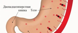
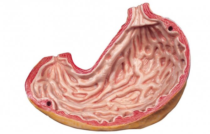
Normally, the gastric mucosa has a light pink color, which becomes brighter closer to the pyloric region.
In some patients they have a yellowish tint, which is not a pathology. During examination, the epithelium reflects the light of the endoscope, so it appears shiny. Numerous folds of the mucosa have a thickness of 6-10 mm.
Their size gradually increases closer to the antrum. When air is introduced into the stomach cavity, the folds of the mucous membrane are smoothed out, and this allows you to examine the entire surface.
If the diagnostician notes that the gastric mucosa is hyperemic, what does this mean? External signs of hyperemia are redness and swelling of the folds of the stomach. The color change is due to blood flow.
The mucous and submucosal layers of the wall have a branched capillary network, with numerous anastomoses between them. Therefore, an increase in blood inflow and a decrease in blood outflow causes the filling of capillaries, which shine through the epithelial layer, changing the color of the mucous membrane.
The chronic form of gastritis often does not have significant manifestations and manifests itself only in a slight decrease in the overall quality of life of the patient. Mild forms of the disease are characterized by stool disorders, and severe forms are characterized by anemia, frequent passage of gas, and bad breath.
Depending on the state of the acidic environment of the stomach, the symptoms of the disease differ slightly. So, with increased acidity, the following signs are noted:
- pain in the middle of the hypochondrium that goes away after eating
- diarrhea
- heartburn after eating acidic foods
- frequent belching
Reflux esophagitis
Gastritis
An accurately identified form of gastritis is the key to successful treatment, which is comprehensive. Neglect of the pathology and failure to comply with doctor’s instructions complicates the treatment of gastritis. For this reason, the outcome of the disease depends only on the patient’s desire to quickly eliminate the stomach problem. A twice-a-year examination by a gastroenterologist will relieve sudden onset pathologies.
Causes of the disease
The most common cause is infection with the bacterium Helicobacter pylori. But it is worth noting that bacteria develop only in a fertile environment, which can arise due to poor nutrition, other diseases, and abuse of bad habits.
The main reasons are as follows:
- cytomegalovirus or candidiasis;
- bacteria Helicobacter pylori;
- the predominance of spicy and hot foods in the diet;
- bad habits;
- infection with worms;
- roughage;
- a large amount of salt;
- drinking carbonated drinks;
- harmful working conditions associated with toxic substances;
- use of certain medications;
- prolonged stress;
- pathological processes in blood vessels;
- taking NSAIDs;
- autoimmune diseases.
If antral gastritis is treated incorrectly or untimely, a narrowing of the lumen of the stomach may develop! This will lead to impaired peristalsis and the appearance of scars.
Classification of mucosal hyperemia
There are passive hyperemia, which is characterized by excessive blood flow, and active (when the flow of blood from the wall of the organ is impaired). The passive type of hyperemic mucosa is a violation of the venous circulation in the organ. The active form is arterial hyperemia.
In the first case, the organ continues to be damaged as a result of oxygen deficiency. Staying active promotes recovery.
In addition, hyperemia can be focal or diffuse depending on the location.
Diagnostics
Having looked at the statistics, we can conclude that almost 90% of people need consultation with a gastroenterologist. To make a correct diagnosis, a specialist prescribes an examination, which is divided into laboratory and instrumental diagnostics.
Laboratory methods include: studies of gastric juice, blood, urine and feces. With their help, you can determine the secretory function, bacterial composition of the gastrointestinal tract, enzyme activity and other important functions. But without instrumental methods, the analysis results are uninformative.
Laboratory methods include: studies of gastric juice, blood, urine and feces. With their help, you can determine the secretory function, bacterial composition of the gastrointestinal tract, enzyme activity and other important functions. But without instrumental methods, the analysis results are uninformative.
The gastric mucosa is focally hyperemic, there is a coating with whitish foamy mucus on the surfaces of the organ in “mucus lakes”, the folds are compacted and are not completely smoothed out with the help of air.
To detect the disease - even if there are almost no problems with the stomach - make an appointment with a gastroenterologist. Gastroscopy is an excellent diagnostic option. Diagnostics involves a procedure carried out by a probe, camera and inspection optics. Using this method, you can assess the condition of organs, take a tissue biopsy, find out the diagnosis and prescribe therapy.
A gastroenterologist will help diagnose the problem. He first examines the patient and collects anamnesis.
After a medical examination, a gastroscopy is performed. It is performed using a special device - an endoscope. It is equipped with viewing optics and a camera.
This diagnosis is an unpleasant and painful procedure, but it allows you to accurately determine the condition of the organ, identify the causes of hyperemia, thanks to which the doctor prescribes the appropriate treatment tactics. In addition, using this method, a biopsy is performed, i.e., tissue is taken for examination.
For preventive purposes, as well as at the slightest suspicion of gastrointestinal diseases, you should seek help from a gastroenterologist. The most accurate and reliable research method is gastroscopy. This procedure involves attaching a camera and optics to the probe, as well as the use of other auxiliary instruments that are inserted deep into the esophagus and stomach.
Gastroscopy allows you to accurately determine the condition of the internal organs of the gastrointestinal tract, identify pathological changes and, if necessary, take a tissue sample for further study.
A doctor can easily notice pathological changes in the epithelium, since in a healthy state they are characterized by transparent mucus, as well as a shiny pink surface. Diagnosis is made on an empty stomach, which allows you to notice the slightest changes not only in the epithelium itself, but also in its thickness.
Gastric hyperemia is characterized by redness of the mucous membrane and indicates the onset of the development of many gastrointestinal diseases. Early diagnosis allows you to determine the cause of its development, as well as eliminate it as quickly and effectively as possible.
Stages and symptoms of the malignant process
The development of a cancerous tumor occurs gradually, over years, and there are 4 clinical stages, the criteria of which are the degree of damage to the stomach and other organs by the tumor. In the modern international classification, symbols are used to indicate individual characteristics of the tumor:
T –
means the tumor itself, its size;
N – damage to lymph nodes by metastases (nodulus – node);
M – presence of distant metastases in organs, bones (metastasis);
The following gradation of these characteristics is accepted.
T (tumor size)
T1 – tumor within the mucous membrane;
T2 – tumor grows into the submucosal and muscular layer;
T 3 – germination of the entire thickness of the gastric wall;
T4 – spread beyond the stomach to neighboring organs.
N (lymph nodes)
N 0 – no metastases were detected in the lymph nodes;
N1 – single metastases in the nearest lymph nodes;
N2 – multiple lesions (up to 15) of lymph nodes;
N 3 – damage to more than 15 lymph nodes, metastases to distant nodes.
M (distant metastases)
M 0 – no metastases detected;
M1 – distant metastases were detected (in organs, spine, bones of extremities).
The G indicator is also used - the degree of malignancy, aggressiveness of cancer (gradus). It can be low (G1), medium (G2) and high (G3), determined histologically by the nature of the cells.
Clinical symptoms of gastric antral cancer depend on the age of the tumor and increase as it grows and spreads. It is characteristic that the very initial 1st stage in most cases does not manifest itself in anything. Only when the tumor spreads to the muscle layer and beyond do symptoms appear:
- feeling of fullness in the stomach;
- nausea, belching with a putrid odor, vomiting of eaten food;
- heartburn;
- bloating;
- pain in the epigastrium, in the presence of metastases - pain in the liver, spine, bones, joints;
- loss of appetite, sudden weight loss, pallor, anemic skin (late symptoms with the development of cancer intoxication).
Symptoms of impaired gastric emptying prevail; gastric bleeding may occur - vomiting with blood, black tarry stools.
Symptoms of gastritis development
In such situations, additional examination is required. Hyperemia is often observed in patients with unstable mental health and under stressful conditions.
It is very difficult to diagnose chronic gastritis, the symptoms of which cannot always be recognized immediately. Many patients do not pay attention to the unpleasant sensations, thereby missing an important point.
But according to statistics, every 5 inhabitants of the planet suffer from a chronic form of this insidious disease. The worst thing is that peptic ulcers or stomach cancer often develop against the background of gastritis.
Difficulty of diagnosis
Gastritis is divided into several types and forms of the inflammatory process of the inner lining of the stomach walls. Gastric juice plays an important role in diagnosing the problem. The course of the disease, therapy and symptoms depend on the level of acidity. There is gastritis with high or low acidity.
Providing assistance with hyperemia of the gastric mucosa
If unpleasant symptoms appear in the stomach area due to hyperemia of its mucous membrane, it is necessary to contact a specialist as soon as possible to prescribe the correct treatment. But if you can’t see a doctor right away, you can temporarily use a few simple tips.
Ensure complete rest by taking a lying position. Drink a glass of clean, cool water. Take No-shpa or another antispasmodic drug. Apply a heating pad with ice to the epigastric area. Follow a strict diet by reviewing your diet.
Manifestation of the disease
In gastroenterology, mucosal hyperemia is associated with stomach diseases such as gastritis and peptic ulcers. In various forms of gastritis, in addition to focal hyperemia, the following signs are observed:
- Spicy. It is characterized by severe hyperemia and swelling of the folds, petechiae, erosions, and copious amounts of thick mucus.
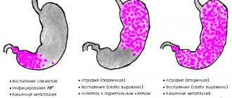
- Chronic. The mucous membrane is pale, dull, grayish in color. Sometimes there are thinned areas (atrophy) with translucent vessels. This is the so-called false hyperemia.
- Superficial gastritis is characterized by diffuse hyperemia, the formation of foamy white mucus, swelling of the folds that do not level out when inflated. Submucosal hemorrhages are sometimes observed.
- Hypertrophic gastritis is characterized by thickening and pronounced diffuse hyperemia of the folds; they acquire a cherry color. Proliferative processes (nodules, warts) are revealed on the surface.
Hyperemia is also present in other forms of gastritis (phlegmonous, necrotic), as well as ulcers. It indicates an inflammatory process. When infected with Helicobacter Pylori, hyperemic manifestations are more pronounced.
Diagnostic methods
Hyperemic changes can only be diagnosed using endoscopy. For diagnosis, fibrogastroduodenoscopy or endoscopic video capsule is used. Other studies (ultrasound, radiography, CT, MRI) can visually determine the appearance of the inner layer only indirectly, by identifying swelling of the mucous membrane.
Pathology manifests itself for two reasons:
- Poor blood removal from the stomach (venous or passive).
- Large blood flow (arterial or active).
In the first case, further recovery is possible, but the passive form affects tissues due to a lack of oxygen.
Gastric hyperemia is characterized by a high blood content in each vessel of all linings of the stomach. In its normal state, it has a pinkish color and a reflective layer of epithelium, which can be seen during endoscopy. The thickness of the folds is 0.5-0.8 cm. When air is supplied to the stomach, they are smoothed out.
Arterial hyperemia develops when in each artery and even in small arterioles that carry blood to the stomach, an increased flow of blood is detected in each banded part of the organ. Various injuries to the smooth muscle of the vessel are characterized by strong blood filling, so the gastric mucosa turns red.
Based on the surface of the gastric mucosa, an experienced specialist can determine the type of disease:
- Superficial gastritis indicates a moderately hyperemic pathology. In this case, there is a white mucous foam on the surface of the stomach, and the folds have tortuous thickenings and do not straighten when air enters.
- In an atrophic state, the mucous membrane seems to become thinner, and upon examination a reddish vascular pattern is noticeable.
- With fibrous gastritis, purulent lesions are visible, since the causative agents of the disease are infections (scarlet fever, measles). Possible vomiting of blood.
- The phlegmous type of pathology indicates only focal areas that have been affected, for example, a fish bone.
The stomach lining becomes red and swollen because the blood vessels in the walls of the organ become overfilled with blood. It is not for nothing that in the old days this condition was called “plethora.”
The first type is called venous or passive hyperemia, the second - arterial or active. There is a significant difference between active and passive hyperemia.
Only active action leads to tissue healing, while passive action, on the contrary, contributes to further damage to the organ due to lack of oxygen in the tissues.
The gastric mucosa becomes hyperemic in most diseases of the gastrointestinal tract.
Based on the condition of the mucous membrane and the location of redness and swelling, the type of disease can be determined.
Most often, hyperemia is diagnosed as one of the types of gastritis, but it can be a symptom of duodenitis, stomach ulcers, or diseases of organs that are not related to the gastrointestinal tract at all.
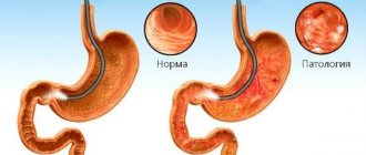
Normally, the gastric mucosa should be pink, shiny, and well reflect the light of the endoscope.
The folds of healthy mucosa are 5–8 mm thick; when air is blown in, they straighten well, allowing the doctor to view all parts of the organ through an endoscope.
Hyperemic gastric lining occurs in several varieties. The type of hyperemia determines the diagnosis of the disease.
With superficial gastritis, hyperemia reaches a moderate degree. The inflammatory process can cover a separate area or become widespread. During the acute course of the disease, the endoscope reveals white foam, the folds of the organ look thicker than normal. When gas is injected, it is not possible to completely achieve a smooth inner wall.
Atrophic gastritis is characterized by focal thinning of the membrane. The vascular pattern in this place is clearly visible, the areas of the mucosa around the atrophic zone look paler.
If the hyperemic gastric mucosa is accompanied by the release of purulent masses, such gastritis has a fibrous form. The disease rarely has independent factors of genesis; in most cases, scarlet fever or measles have consequences in the form of hyperemia of the mucous membrane, followed by vomiting with blood.
This is how areas of dead mucous membrane with pus are rejected and is accompanied by intense pain.
Phlegmous gastritis is usually called hyperemia of an area of the mucous membrane that has been subjected to injury or sexually transmitted infection.
Alkali or acid in the stomach causes deep damage to many layers of the digestive organ. Necrotic areas are not the worst option for the development of necrotic gastritis. It is worse if provoking factors cause perforation of the walls of the organ, pouring its contents into the abdominal space and causing peritonitis.
General information
Focal hyperemia is the overflow of blood vessels with biological fluid, which leads to their expansion and swelling. Normally, the gastric mucosa is pale pink in color. As the disease progresses, its color changes, swelling and redness of the gastric folds appears. The affected area sometimes takes on a marbled or bluish tint. As a result, the functioning of the organ is disrupted, causing the entire digestive system to suffer.
Reasons for development
Hyperemia of the gastric mucosa is not an independent pathology, but is a clinical manifestation of gastric diseases. The following gastrointestinal diseases can provoke a pathological condition:
- Gastritis is inflammation of the inner (mucous) lining of the stomach. The form of the disease is determined taking into account the degree of damage. Acute inflammation is accompanied by pain, a feeling of heaviness, belching, drooling and other symptoms. As a rule, the chronic course of the disease does not have a pronounced clinical picture. If the mucous membrane is diffusely hyperemic, then we are talking about superficial gastritis.
- Reflux esophagitis is a chronic pathology characterized by pathological reflux of gastric contents into the esophagus. Due to stagnation of food in the stomach, the internal membranes of the organ are subject to constant stress, which leads to a change in their structure. Chest pain, angina-type pain syndrome are specific signs of the disease.
- Bulbit is a catarrhal or erosive inflammation of the bulbar part of the duodenum. In most cases, the etiological factor of bulbitis is infection with Helicobacter. The disease may occur without clinical symptoms. Nausea, vomiting, bloating, unpleasant taste in the mouth, white coating on the tongue are the main symptoms of bulbitis.
- Duodenitis is an inflammatory disease of the duodenum, in which its mucous membrane is most severely affected. Constant nausea, yellowing of the skin, rumbling in the stomach, flatulence, loss of appetite are symptoms of the disease. Duodenitis is often combined with gastritis.
- Peptic ulcer disease is a chronic polyetiological pathology that occurs with the formation of ulcerative lesions in the stomach, a tendency to progression and the formation of complications. The severity of clinical manifestations depends on the location and degree of damage to the inner membrane.
Attention! Peptic ulcer disease is extremely dangerous, as it can lead to perforation, stenosis, and bleeding.
The following unfavorable factors can contribute to the appearance of swollen and reddened areas of the mucous membrane:
- taking certain medications (non-steroidal anti-inflammatory drugs, corticosteroids);
- mechanical damage to the stomach;
- poor diet (eating large amounts of junk food);
- infectious pathologies;
- presence of the bacterium Helicobacter Pylori;
- renal failure;
- chronic intoxication;
- bad habits (smoking, drinking alcohol);
- prolonged stress, depression;
- hereditary predisposition.
In some cases, hyperemic gastric mucosa is a consequence of inflammation of the walls of the organ.
Prevention
It is also necessary to exercise and not overwork. This will help boost immunity and ensure protection and rapid restoration of the mucous membrane.
To prevent the development of further pathological process, it is necessary to undergo a comprehensive examination and establish the exact cause, which will help to choose the right treatment.
Following some rules will help prevent stomach diseases:
- Eating the right foods.
- Rejection of bad habits.
- Take medications only as prescribed by a doctor.
- Weight control.
It is also recommended to undergo gastroscopy twice a year to prevent problems with the gastrointestinal tract. The onset of hyperemia indicates a stomach disease.
There is probably no person who would like to experience any problems with the gastrointestinal tract, giving up their favorite foods, taking many medications and undergoing unpleasant examination procedures. To avoid this, you need to change your usual lifestyle a little and follow some simple rules.
The key to favorable progress and complete cure of diseases, and therefore of symptoms, is an accurate diagnosis with determination of the root cause of its occurrence. Treatment becomes more complicated if the doctor’s recommendations are not followed. Therefore, it is important to undergo a physical examination and gastroscopy with a gastroenetrologist twice a year, which will avoid sudden problems with parts of the stomach.
In addition, it is important to eat right, get rid of bad habits, avoid stress, and not abuse gastro-soluble medications.
To prevent the development of hyperemia of the gastric wall, you need to adhere to the basic rules of prevention. First of all, it is important that the diet is balanced and rational. Therefore, it is necessary to include healthy foods in your diet and avoid junk food.
Forecast and preventive measures
Oncological diseases, in principle, do not have a favorable prognosis for health and life. It depends on the stage of the tumor, the quality of treatment, the age and health of the patient. In particular, for antral cancer, 5-year survival statistics are as follows:
- with stage 1, 80-85% of patients survive;
- with stage 2 – 55-60%;
- with stage 3 – up to 25%;
- with stage 4 – no more than 3-5%.

Unfortunately, in the vast majority of patients this diagnosis is detected at stages 3-4. In this regard, preventive measures are needed aimed at reducing the risk of developing cancer and possibly early diagnosis, detection and treatment of precancerous conditions. Such measures include:
- Quitting smoking and drinking alcohol.
- Normalization of nutrition, consumption of healthy, healthy foods.
- Regular medical examinations.
- Timely treatment of existing chronic diseases.
Some people are skeptical about cancer prevention, and this is a big misconception. Practice shows that a dangerous disease can be prevented to a large extent by eliminating risk factors that create favorable conditions for its development.
Treatment methods
It has been proven that treatment at home can give fairly good results. All that is required of you is a responsible and serious approach to treatment. The most effective and efficient methods of treating inflammation are the recipes given below.
Traditional method 1. Prepare a water infusion of lettuce leaves. Drink one glass of the resulting product daily.
Folk method 2. Prepare a decoction of sea buckthorn berries, add a little honey to it and drink a glass of the resulting product every time you are going to have a snack.
Folk method 3 for symptoms of inflammation of the mucous membrane. Eat about 8 grams of propolis daily on an empty stomach. Chew it thoroughly before swallowing.
Folk method 4. Pour a little dried thyme herb into a liter of white wine, wait one week, then bring the mixture to a boil and strain. If you are diagnosed with inflammation of the stomach, drink a glass of the resulting product per day, after dividing the daily dose of the drug into 4 equal doses.
Traditional method 5 for the treatment of inflammation of the mucous membrane. Pour a glass of oatmeal (oat flour) with a glass of water and bring the resulting mixture to a boil. Try to drink as much of this jelly as possible during the day. To improve the taste, you can add a little cranberry juice to it.
Since the gastric mucosa can become hyperemic for various reasons, this means that drug treatment is not always required. Sometimes it is enough to eliminate the impact of adverse factors on the body.
Treatment is carried out in accordance with the diagnosis. Appointed:
- agents that protect the mucous membrane;
- antibacterial drugs;
- medicinal substances that normalize the acidity of gastric juice;
- vitamins, etc.
In most cases, hyperemia of the gastric mucosa is not treated, since in order to combat the pathological process it is necessary to eliminate the disorders in a natural way. Usually the body fights the problem on its own. Hyperemia accelerates metabolism, resulting in the restoration of damaged tissue. However, there are cases when doctors artificially increase blood flow.
Often redness and swelling of the gastric mucosa indicates gastritis, which requires long-term therapy.
Treatment includes:
- dietary nutrition;
- taking medications;
- antibiotics for infection;
- rejection of bad habits.
The problem of how to treat mucosal atrophy depends on the predominant aggressive action, the identified cause of the process, and the residual ability to recover (reparation). Given the absence of severe symptoms, patients are often treated on an outpatient basis. Mandatory recommendations include: regimen and diet.
It is not recommended to engage in strenuous sports; it is necessary to reduce physical activity to moderate. It is required to stop smoking and drinking alcoholic beverages, including beer. It is prohibited to take any medications without permission, including those for headaches and flu.
Diet Requirements
The patient's diet includes choosing foods that do not damage or irritate the gastric mucosa. Therefore, it is strictly prohibited:
- fried, smoked, salted and pickled dishes;
- strong tea, coffee, sparkling water;
- ice cream, whole milk;
- confectionery, fresh baked goods;
- spices, sauces, canned food;
- legumes
The patient is advised to maintain meals in small, frequent meals. Use stewed, boiled, steamed, baked dishes. In case of pain, it is advised to switch to semi-liquid pureed food (meatballs, low-fat broths, oatmeal with water, jelly) for several days.
In many cases, hyperemia is not treated, as it is considered a sign that the body is effectively fighting damage through self-regeneration. Hyperemia helps accelerate metabolic processes, which triggers self-healing and tissue healing.
But such a diagnosis is considered the norm when it comes to arterial hyperemia. Sometimes doctors artificially induce blood flow to stimulate healing.
But more often, redness of the epithelium indicates gastritis, which is treated over a long period of time and comprehensively: diet, medication (for example, antibiotics for Helicobacter pylori infection). For gastric pathologies, the use of home remedies is indicated - herbal remedies, honey, a special diet. Diet therapy for hyperemic mucosa is based on therapeutic nutrition according to the principle of Professor Pevzner.
Treatment
If antral gastritis has been detected, it is necessary to prescribe therapy that will be designed to reduce the level of acidity, destroy pathogenic flora, eliminate all associated diseases, relieve inflammation and restore the mucous membrane and glands.
Treatment can take place both in a hospital and at home. The most important thing is that the treatment takes place in a complex manner. It must include both medications and traditional methods when following a diet.
Therapy is divided into two main stages - the initial goal is to eliminate the bacteria causing the disease. In the second, restoration of the organ and its functions.
Medicines are used at the initial stage in the following areas:
- antibiotics and antibacterial agents;
- reduction of pain;
- lowering acidity and blocking receptors;
- reduction of side effects.
At the next stage, preventive methods and methods of healing the body are added to medications.
Antibacterial therapy
It is important to carry out this type of treatment on an outpatient basis. The full course is about two weeks. Medicines of the group of proton pump inhibitors, prokinetics and receptor blockers are used. The most effective was considered to be a triple scheme - the use of an inhibitor and two antibacterial agents. If there is no effect, a four-drug approach is used.
Secretory therapy
Helicobacter pylori bacterium
Due to the bacterium Helicobacter pylori, the composition of gastric juice undergoes changes, accompanied by an increase in acidity. Secretory therapy is designed to reduce the effect of acidic gastric juice on the mucous membrane and its corrosion. Drugs in this group include Almagel, omeprazole, Rennie and others. Digestion is adjusted by using enzymatic preparations - panzinorm, festal. In case of disruption of the pancreas, Creon, Mezim and Pancreatin are prescribed.
Second stage of therapy
At this stage, they try to restore the functions of the stomach. To protect the mucous membrane, enveloping drugs are used - sea buckthorn oil, retabolin, sucralfate. Actovegin and similar drugs have regenerative properties - they help restore the mucous membrane.
Vitamin therapy is used as prevention and general health improvement of the body. Vitamin U helps correct the secretory function of the stomach. Vitamin B5 helps restore mucosal cells and has a good effect on the digestive process.
Traditional methods of treatment, a gentle diet, a healthy lifestyle, and physiotherapy will also not be superfluous.
Features and Recommendations
If hyperemic mucosa is detected, the patient is advised to follow a diet. Substances that irritate the mucous membrane should be excluded from the diet: spicy, salty, sour, fatty foods, smoked meats and marinades, alcohol, strong coffee. Fried foods are not recommended. For heat treatment, food should be boiled or steamed.
The main cause of exacerbation of pancreatitis is non-compliance with the diet prescribed by the doctor. Establishing a proper diet for pancreatic disease during the period of deterioration will help relieve painful symptoms. The tablet therapy process will be much more effective.
Do I need to follow a diet for pancreatitis?
The main component of treatment for exacerbation of pancreatitis is diet. When the acute phase of the disease develops, the patient's body needs complete rest. It is necessary to provide the pancreas with a significant reduction in its excretory function.
Nutrition for gastric hyperemia
Possible complications and prognosis
After the underlying disease of the stomach is cured, a symptom such as redness of the mucous membrane goes away on its own.
In addition, any form of gastritis can lead to peptic ulcers, which can even be fatal if severe.
If you have stomach problems, the condition of your nails, skin and hair worsens.
To avoid the development of undesirable consequences, it is important to promptly diagnose diseases that are accompanied by gastric hyperemia and begin timely treatment. Therefore, if there are any signs of diseases of the digestive organs, you must consult a gastroenterologist.
Types of illness
Antral gastritis comes in several forms:
- Superficial - occurs in the outer layer of the gastric mucosa;
- Catarrhal - superficial gastritis, developed at the very bottom of the antrum;
- Focal - with it, atrophied zones and damaged epithelium are clearly expressed;
- Erosive - serious damage to the mucous membrane, provoking the appearance of erosions and the development of ulcers;
- Erythematous - can develop into an erosive form, characterized by pink rashes on the mucous membrane;
- Hyperplastic - swelling of the mucous membrane, leading to the appearance of cysts;
- Granular - hyperplastic form with cysts up to 2 centimeters in size;
- Follicular - a rather rare form in which the glands that synthesize bicarbonates are clogged;
- Atrophic antral gastritis is a precancerous condition that leads to gastric insufficiency.
There is also mixed gastritis, when several of the above forms can be observed at once.
The disease can be divided into forms and according to its severity:
- Stage 1 - minor changes in cells in the tissues of the organ;
- Stage 2 - atrophy and infiltration of medium size;
- Stage 3 - high level of atrophy and infiltration.