Analysis of stool for Helicobacter pylori: preparation and interpretation of indicators – In the abdomen
Analysis of stool for Helicobacter pylori is reliable evidence of the presence in the human body of a bacterium that causes ulcers and many other diseases of the human digestive tract. Such an analysis is direct evidence if either Helicobacter itself or its DNA is detected. The reliability of the analysis is the highest of all known.
How to take the test correctly?
The result depends on the correct preparation and collection of material. No human presence is required; it is enough to deliver the correctly collected material on time.
Preparing for the test
When preparing, you must strictly follow the following rules:
- donate stool for analysis no earlier than 1 month after the last dose of antibiotics;
- during the 3 days preceding the collection of material, you should not consume coloring products (tea, coffee, red and orange fruits and vegetables, turmeric, curry, soy sauce),
- exclude coarse fiber - cabbage, radish, radish, pearl barley, bran, beets;
- You should not take medications that enhance intestinal motor function - Lactulose, Motilium and the like;
- give up alcohol;
- do not take antacids (Almagel and the like);
- collect material during the first week after the onset of symptoms of the disease (then the concentration of Helicobacter in feces decreases).
3 days are enough for preparation. At this time, it is advisable to stop taking all medications (if this is not possible, tell your doctor the names and dosage).
You need to reduce your consumption of fish and meat, steam or boil them.
The best food for this time is porridge and fermented milk products, vegetable and fruit purees, weak soups, neutral natural juices (apple, white grape), compotes, fruit drinks, weak tea, rosehip decoction.
Preparing containers
As a last resort, you can use a glass jar with a well-fitting lid (baby food). The jar needs to be washed well and then boiled along with a lid and a spoon or other object that will be used to collect feces. If bacteria remains on the dishes, the analysis will be incorrect.
Collection of material
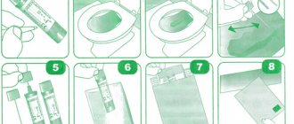
You cannot collect material from the toilet or diaper. The toilet should be covered with a clean plastic bag, and instead of a diaper (for a child or a bedridden patient), an oilcloth should be laid. It is allowed to take material from a clean pot.
The container is filled no more than one third, close the lid tightly. Attach the directions to the container or write on the label (legibly) the patient's first and last name, year of birth.
How and for how long can the material be stored?
The container with the material is delivered to the laboratory as early as possible, optimally on the day of collection. If there is no possibility of urgent delivery, storage in the refrigerator for no more than 2 days at a temperature not exceeding +4 °C is allowed. If longer storage is necessary, the material is frozen once at a temperature of -20 °C.
Methods for analyzing and deciphering indicators
Biological material can be studied in different ways.
| Method | Dates | Decoding the results |
| PCR | 1 working day |
|
| Bakposev | from 6 to 12 days |
|
| ELISA | Material – venous blood, during the working day, urgently within 2 hours |
|
Other ways to identify Helicobacter
During the examination, several methods are usually used to finally verify the presence or absence of Helicobacter:
- Breath test. You exhale into the prepared bag, then give medical radioactive carbon to drink, after 10 or 30 minutes you need to exhale into another bag. Detects the presence of Helicobacter in very small quantities.
- Rapid urease test of gastric biopsy. It is based on the fact that the bacterium secretes urease, which breaks down urea into ammonia and CO2. During an endoscopic examination, 4 biopsies are taken from different parts of the stomach and placed in the cells of the CLO test. The more Helicobacter, the faster the color of the cell changes.
- Helicobacter antigen in feces. Particles of the bacterial wall react with the monoclonal antibody, which changes the color of the indicator. Test cassettes are available for home use. According to the instructions, feces are mixed with a solvent (a test tube with it is included), a few drops are applied to the cassette window. Two stripes are positive, one is negative. Additionally, you can find out from the marking whether the test is valid.
- Histological examination of the biopsy specimen. The biopsy obtained during FGDS (5 samples) is stained using various methods, the sensitivity of the analysis reaches 99%.
- Western blot. It is carried out in those who have antibodies to Helicobacter using the ELISA method (described above). The number of antibodies of each class (A, G, M) is determined.
The range of diagnostic tests depends on the equipment of the medical institution. Histological examination is considered the “gold standard”. However, a positive result from at least two different tests is considered sufficient to confirm the presence of the bacterium.
What to do if the test is positive?
Visit a gastroenterologist as soon as possible to find out whether the bacteria need to be eradicated (removed from the body). This is an ambiguous process, there are contraindications, consultation with a doctor is necessary.
After assessing the level of health, the doctor selects one of the regimens approved by the international community. Depending on the characteristics of the pathological process, drug combinations of drugs from the following groups are used:
- proton pump inhibitors - Omeprazole, Rabeprazole and others;
- Clarithromycin is a macrolide antibiotic;
- amoxicillin – semi-synthetic penicillin;
- Metronidazole is an antiprotozoal drug with antibacterial activity;
- bismuth preparations are anti-ulcer drugs that have a protective effect on the gastric mucosa;
- tetracycline is a broad-spectrum antibiotic.
Helicobacter tests may be repeated during treatment to monitor the effectiveness of treatment.
After the bacteria are detected and removed, ulcers and other mucosal lesions heal completely, and all secondary disorders stop - microflora, synthesis of vitamins and hormones, digestion are restored, and the risk of developing stomach cancer is reduced.
Source:
Stool analysis for Helicobacter pylori: preparation and interpretation
Helicobacter pylori is one of the most dangerous bacteria that provokes the development of various diseases of the stomach and duodenum. To diagnose pathology, not only a blood test is prescribed, but also a stool test.
Helicobacter pylori: causes and symptoms
Helicobacter pylori is a pathogenic bacteria that infects the duodenum and stomach
Helicobacter pylori is a pathogenic bacterium that, when it enters the body, causes the development of helicobacteriosis. It is small in size and has a spiral shape. Producing a large amount of toxins, it affects the stomach and duodenum. The bacterium gets its name from the fact that it is found in the pyloric region of the stomach.
By multiplying, Helicobacter affects all cells of the stomach, which leads to inflammatory processes. This bacterium is resistant to acidic environments and can move along the walls of the stomach using flagella.
The following symptoms are characteristic of Helicobacter pylori infection:
- Heartburn
- Nausea and vomiting
- Smell from the mouth
- Loss of appetite
- Bloating
- Stool disorder
- Discomfort after eating
The listed symptoms are similar to those of gastritis and ulcers, therefore, to identify the pathogen and treat helicobacteriosis, it is necessary to undergo an examination.
Stool analysis: preparation and collection of material
Medical disposable containers for collecting stool for analysis
A stool test is a laboratory test that can detect parasites living in the intestines. Using this analysis, you can accurately determine Helicobacter in the body. The reliability of the results depends on proper preparation and collection of material:
- It is necessary to stop taking antibiotics, laxatives, and rectal suppositories 3-4 days in advance. It is important to notify your doctor about the use of medications.
- The material for analysis must be collected in a clean container. The pharmacy sells a special container for this purpose. It is prohibited to collect material after enemas or taking laxatives, as the results will be unreliable.
- After collection, the material can be stored for 12 hours at a temperature of 2-8 degrees, but for research it is better to submit it 4-5 hours after the bowel movement.
- If stool collection is performed after antibiotic therapy for helicobacteriosis, then this should be done only a month after the end of taking antibiotics.
: What is hydrocephalus and why is it dangerous?
In the laboratory, the resulting material is examined using the PCR method. This is a molecular genetic diagnosis that allows you to identify Helicobacter even a small fragment of bacterial DNA in the collected material. The study is carried out under certain conditions and temperature conditions.
More information about Helicobacter pylori can be found in the video:
Read: What does dark-colored stool indicate?
To detect Helicobacter pylori infection, other methods can be used: cultural and immunological analysis. The cultural method is usually understood as bacteriological inoculation of the material under study. It is placed in a special environment for bacterial growth.
Usually it is about 10 days. Then the type of bacterium and its ability to enter into biochemical reactions are examined under a microscope. This method allows you to determine the sensitivity of the identified bacteria to antibiotics, which allows you to prescribe correct and adequate treatment.
The immunological method involves the adhesion of antibodies to antigens. This method of examination is carried out in case of severe symptoms and before the instrumental examination method. The choice of a specific method is made by the doctor, taking into account various factors.
Decoding stool analysis
Stool bacterial culture analysis involves a qualitative and quantitative assessment of microorganisms in the test environment. There are 4 degrees of bacterial growth:
- 1st degree. In a favorable liquid environment, the growth of the identified bacteria is weak, and in a solid environment it is not observed at all.
- 2nd degree. The reproduction and growth of a certain type of microflora reaches up to 10 colonies.
- 3rd degree. It is distinguished by significant growth of up to 100 colonies in solid media.
- 4th degree. The growth of the bacterium is very high and exceeds 100 colonies.
The third and fourth degrees indicate an inflammatory process provoked by a specific type of bacteria.
The presence of Helicobacter in feces does not indicate a stomach ulcer or other diseases of the gastrointestinal tract, but only determines the presence of a pathogenic bacterium. If, according to the results of the analysis, the gastric mucosa is colonized with Helicobacter, then in many cases this is accompanied by pathologies such as ulcers, gastritis, irritable bowel syndrome, carcinogenic tumor, pancreatitis, etc.
Laboratory tests alone are not enough to make an accurate diagnosis. The examination must be comprehensive and include a biopsy and histological examination.
Helicobacter during pregnancy: danger to the fetus and treatment methods
The pathogenic bacterium does not affect the course of pregnancy and the condition of the child
During pregnancy, the gastrointestinal tract is subject to heavy load. In this case, heartburn, pain in the epigastric region, etc. are very often observed. The risk of developing an ulcer is very high, so the expectant mother should be examined to identify this infection in order to avoid possible violations.
It is better to carry out the examination in the first months, since diagnosis becomes more difficult as the period increases. Non-invasive research methods are used for diagnosis.
Source: https://carmenta-med.ru/lechenie/analiz-kala-na-helikobakter-pilori-podgotovka-i-rasshifrovka-pokazatelej.html
Detection of Helicobacter pylori in stool
This method of studying is quite comfortable for most people, since it does not even require the direct presence of a person at the reception. The positive aspects also include the fact that the analysis is carried out without any harm to the human body. That is why this type of analysis is recommended for young children, the elderly, or those who have recently undergone surgery. Stool donation is carried out at home after several days of certain restrictions. What are these restrictions? Do not eat:
- products with a large amount of coloring substances;
- foods rich in dietary fiber;
- many inorganic salts;
- pharmaceutical preparations that enhance intestinal motility.
Blood test for Helicobacter pylori: deciphering the indicators
Helicobacter pylori is a gram-negative bacterium that inhabits the gastrointestinal tract and can exist there without air. Once in the body, the bacterium settles in the stomach. It belongs to the only species that is not exposed to gastric juice.
Transmitted in the following ways:
- through saliva;
- through mucous secretions;
- with dirt;
- through unwashed foods.
The main cause of Helicobacter pylori infection is considered to be weak immunity throughout the body and locally in the digestive organs.
Helicobacter does not always lead to devastating consequences. In its dormant state it is harmless. Here the degree of expression of the body’s own defenses is of great importance.
A little about the features of Helicobacter
The literal Latin-Greek name Helicobacter pylori (“spiral pyloric”) is associated with the characteristic shape of the bacterium and its maximum residence in the zone of transition from the stomach to the duodenum (pylorus).
With the help of flagella, mobility and the ability to move in the gel-like mucus environment on the inner surface of the stomach are ensured. This is the only microorganism that can live in an acidic environment.
From its discovery in 1875 to the awarding of the Nobel Prize in 2005, 130 years passed. Many scientists have invested their knowledge and experience in studying the unusual infection. She did not grow on nutrient media.
Then, 10 days later, endoscopy showed a connection between the emerging signs of stomach inflammation and the presence of Helicobacter.
Marshall and his colleague Warren did not stop there. They were able to prove the cure of gastritis with a course of Metronidazole and a bismuth preparation, and showed the role of antibiotics in the treatment of gastritis, gastric and duodenal ulcers.
Helicobacter has many aggressive properties caused by its internal composition
Modern research has clarified the conditions for the existence of the microorganism. Helicobacter pylori uses the energy of hydrogen molecules released by intestinal bacteria. Synthesizes enzymes:
- oxidase;
- urease;
- catalase.
In hazardous conditions, it forms a film that protects against the body’s immune reactions. Over time, it changes its shape to spherical (coccal) and in this state is also contagious.
The important point is that it is in the stomach of a person without signs of disease. But if the protective forces fall, it behaves very aggressively, causing inflammation to ulcers and cancerous degeneration. That is why timely detection of traces of Helicobacter pylori in a blood test is so important for human health.
Then, 10 days later, endoscopy showed a connection between the emerging signs of stomach inflammation and the presence of Helicobacter.
- oxidase;
- urease;
- catalase.
When to worry
Testing for Helicobacter pylori is strictly mandatory and regular if it is known that there are ulcerative lesions on the gastrointestinal tract.
The following symptoms indicate serious changes in the functioning of the digestive system:
- Pain while eating. Caused by food stagnation or indigestion due to low fermentation.
- Pain on an empty stomach. They occur when long periods of time pass between meals. The symptom goes away after eating. During a meal, a person seems to feel its movement along the intestinal tract, this is explained by the presence of damaged areas on the gastric mucosa.
- Heartburn is a negative, strong effect of stomach acid on the esophagus, causing irritation. If there are isolated manifestations, there is no reason to worry, but in case of regular discomfort, it is better to undergo an examination.
- Heaviness in the stomach. Occurs after eating. Among the sensations, patients describe feeling as if food could not be digested.
- Nausea. The frequent occurrence of a symptom that is not associated, for example, with pregnancy, also sometimes serves as a signal for analysis.
- The so-called “breakdown” of the stomach. In this case, the person cannot even eat food due to acute painful sensations.
- Discomfort during meals. The main symptoms include aching pain and lack of appetite.
- The appearance of mucus, blood and particles of undigested food in the stool.
Most doctors recommend taking a blood test for antibodies to Helicobacter pylori periodically in addition to undergoing a general medical examination. But it is very rare that any person will consult a doctor unless absolutely necessary. However, the slightest delay in the fight against pathogenic bacteria is fraught with serious complications.
Testing for Helicobacter pylori: interpretation of results and possible deviations
Diagnosis of Helicobacter rulori is very important; it reliably establishes the presence or absence of the bacterium in the human body and allows one to determine treatment tactics if the pathogen has been identified. It is equally important to be able to interpret the results of the surveys.
If an increase in the concentrations of antibodies G, A and M is observed, an aggressive infection process is implied. When the IgG concentration decreases to 2% over six months, we can talk about recovery.
If the IgG type indicator is no longer present during the next examination, this is a signal that Helicobacter pylori has been destroyed. It is better to carry out the analysis 10-12 weeks after treatment. Immunoglobulin type G, when suppressing a bacterial infection, should decrease by 50% by that time.
If antibodies are found, that is, the test shows a positive result, but no symptoms are observed, then passive infection with Helicobacter pylori is detected.
Blood testing is undoubtedly an effective way to detect the presence of infection in the body. As a rule, one analysis is enough for an accurate result. The Helicobacter bacterium is vulnerable to antibacterial effects. And timely treatment is the key to good health.
Source: https://zhivot-info.ru/analiz-na-khelikobakter/
Indications for examination
Despite the fact that Helicobacter pylori is one of the most common human infections (according to some data, more than 70% of the world's population over 40 years of age are carriers of the bacterium), its presence in the body does not always lead to pathological disorders. Most people infected do not show any symptoms. The reasons indicating the need to consult a gastroenterologist are the following:
- aversion to protein (meat) foods;
- feeling of heaviness in the stomach;
- vomit;
- nausea, heartburn, frequent belching;
- pain in the stomach (especially subsiding after eating) and other parts of the abdomen;
- flatulence;
- loss of appetite;
- dysphagia (difficulty swallowing);
- weight loss;
- constipation, diarrhea;
- appearance in vomit or feces.
It is also necessary to undergo tank analysis in the following cases:
- a history of diseases such as gastric and duodenal ulcers, dyspepsia and atrophic gastritis;
- detection of stomach cancer in a blood relative;
- primary detection of Helicobacter pylori in one of the family members living together.
Most often, infection occurs within the family and in closely communicating groups (schools, kindergartens) through household means through mucus and saliva. Crowding, poor hygiene, and sharing utensils increase the risk of infection. In the case of asymptomatic carriage, the impetus for the development of diseases can be such negative factors as smoking, poor diet, alcohol abuse, and stress.
How to take a stool test for Helicobacter pylori. Doctor's recommendations
Helicobacter pylori is one of the most dangerous bacteria for humans. A pathogenic microorganism can provoke serious deviations in the functional state of the digestive tract and the body as a whole. Bacteria of this type are characterized by increased resistance to numerous factors.
The result of therapy directly depends on a correctly designed treatment regimen. To determine the sensitivity of microorganisms to certain types of antibacterial drugs, special fecal tests are used. Each technique has its own advantages and disadvantages. To obtain accurate results, it is recommended to conduct several tests.
Who is recommended to have a stool test tested for Helicobacter?
Helicobacter pylori is present in the body for a long time without symptoms. Signs of disturbances in the functional state of the gastrointestinal tract appear only when bacteria are active. One of the main provoking factors is decreased immunity.
If a patient has certain diseases or individual symptoms, the test procedure for helicobacteriosis becomes mandatory.
Indications for analysis:
- carrying out antibacterial therapy (to monitor treatment results);
- erosions and ulcers of the duodenum or stomach;
- symptoms of dysfunction of the digestive tract (regular constipation or diarrhea, flatulence, pain in various locations);
- genetic predisposition to diseases caused by Helicobacter pylori (malignant tumors of the digestive organs, chronic pathologies of the digestive tract);
- progressive atrophy of the mucous membranes of the stomach;
- regular pain in the stomach after eating food;
- Iron-deficiency anemia;
- a sharp decrease in body weight in combination with other symptoms of abnormalities in the functional state of the digestive tract;
- presence of blood in vomit or feces;
- identified neoplasms and polyps in the digestive system;
- esophageal ulcer or gastroesophageal reflux.
We recommend watching the video:
How to take the test correctly?
Carrying out the procedure for examining stool for the presence of pathogenic bacteria requires following certain rules.
If preparation for analysis or collection of biological material is done incorrectly, the test results may show a false result. Feces have a certain shelf life.
If the material is delivered to the laboratory late, the testing may also become unreliable.
Tactics for determining Helicobacter bacteria using stool analysis
The doctor prescribes a specific test for helicobacteriosis. The specialist takes into account the individual clinical picture of the patient’s health status. You can get tested for Helictobacter pylori yourself if you suspect a functional deviation of the digestive tract.
Molecular diagnostics
Molecular analysis reveals the specific DNA of the bacterium. The code is duplicated on a special device. When the bacteria reaches the desired size, it is compared with ethanol. If parts of the DNA match the original, then the test result is positive (Helictobacter pylori infection has been detected). The technique is characterized by a high degree of information content.
Cultural analysis
Bacterial inoculation (or cultural analysis) belongs to the category of microbiological research methods. During the procedure, biological material is placed in an environment that is most favorable for the proliferation of pathogenic microorganisms. After a week, the stool is examined using a microscope.
To identify Helicobacter pylori, the color of the bacteria and their ability to enter into certain reactions are taken into account.
Features of the technique:
- the procedure allows you to identify a pathological antigen;
- in the process of studying biological material, a sensitivity test to antibacterial drugs is carried out;
- The results of the study allow us to select the most effective treatment regimen.
Determination of Helicobacter pylori antigen in feces by immunological method
The main purpose of immunological tests when identifying Helicobacter pylori is to determine antigen + antibody complexes. Labeled antibodies are used for the study.
When they react with feces, such substances undergo certain changes.
Antibodies are able to detect the presence of not only pathogenic bacteria in the stool, but also their parts, as well as waste products.
Advantages and disadvantages of PCR diagnostics
PCR diagnostics has a number of advantages and disadvantages. Some types of pathogenic bacteria can only be detected using this technique (for example, cytotoxic types of microorganisms). Collecting material for analysis does not imply causing pain or discomfort to the patient. The procedure shows the most reliable results.
Advantages of the technique:
- the procedure allows you to identify various strains of pathogenic bacteria;
- the method has a high degree of sensitivity;
- the analysis is safe for the patient (no medical instruments are used to collect material);
- non-invasiveness and simplicity of the process of collecting biological material;
- high degree of specificity of the test;
- The method allows you to determine not only spiral-shaped, but also coccal types of bacteria.
The high degree of sensitivity of the test is its simultaneous advantage and disadvantage. The analysis accurately determines the presence of bacteria, but if the biological material or container is contaminated, the results of the procedure may be false positive.
To exclude false indicators, you should follow all the rules for collecting stool for testing.
Other disadvantages:
- relatively high price of the procedure;
- inability to determine the sensitivity of bacteria to antibacterial drugs;
- lower sensitivity compared to the procedure for examining a biopsy of the gastric mucosa.
In vitro stool test for Helicobacter pylori antigen
Procedures for studying feces are carried out outside the patient’s body (“in vitro” - “in glass”, “in vitro”). To detect a microbe, such methods are as accurate and effective as possible. However, the procedures have some disadvantages. Some processes occurring in the human body cannot be recreated artificially.
Tests can show the level of sensitivity of bacteria to certain antibiotics, but such tests do not provide 100% information.
Decoding the results
| Procedure | Confidence level of results | Decoding the research results |
| PCR | High | Positive – helictobacteriosis has been detected; negative – no type of Helicobacter pylori was detected |
| Bacteriological analysis for sensitivity to antibacterial drugs | Low – for antigen detectionHigh – for sensitivity determination | The number of bacteria is 0 – the result is negative, no bacteria are detected; indicators greater than 0 – helictobacteriosis is determined; R – resistance to a specific antibiotic; I – moderate degree of sensitivity to the antibiotic; S – high degree of sensitivity to antibiotic |
| Immunological methods | Low | Positive – helictobacteriosis has been detected; negative – no pathogenic bacteria have been identified |
Results
- there are several ways to determine Helicobacter in stool using a laboratory method;
- each analysis option has its own advantages and disadvantages;
- To obtain the most accurate results, it is recommended to take several tests of different types;
- feces for analysis are collected taking into account a number of rules;
- a few days before the procedure, coarse fiber, fried, fatty foods, hot spices and seasonings, alcohol and any orange or red fruits are excluded from the diet;
- the container for feces must be sterile (pharmacy containers are the best option).
on the topic: Why is the Helicobacter pylori bacterium dangerous?
Bibliography
- Descriptive epidemiology of H. pylori infection in patients with dyspepsia NV Bakulina, V Simanenkov, IG Bakulin – 2017
- Inflammatory bowel diseases. Clinical Guide Ed. D.J. Stein, R. Shaker; Per. from English; Ed. I.L. Khalifa 2019
- Doctor. Gastroenterology and hepatology Ed. G.L. Vyshkovsky 2017
Source: https://gastrocure.net/bolezni/khelikobakter/analiz-kala-na-khelikobakter.html
Laboratory immunological method for analyzing Helicobacter pylori
This analysis involves establishing, using special reactions, the presence of immunoglobulins, that is, antibodies, in the blood. They are formed as a response to objects foreign to the body, then combine with the bacterium itself and neutralize it. Blood plasma for study is taken on an empty stomach in the morning. Of course, not everyone has the opportunity to visit a medical institution in the early hours; in this case, you can get tested later, but you must not eat 5 hours before going to the hospital.
What does the presence of occult blood in stool mean?
A small amount of blood in the stool is a normal phenomenon characteristic of the vital processes of the human body . The standard level of blood discharge is no more than 2 mg per 1 g of feces; exceeding this indicator means that hidden blood is present in the stool, the source of which must be further determined.
A positive stool occult blood test indicates the presence of the following diseases:
- intestinal tuberculosis;
- ulcerative exacerbation;
- Crohn's disease;
- polyps;
- malignant and benign tumors in the stomach or intestines;
- ulcerative collitis;
- helminthic infestations;
- haemorrhoids.
Subsequent analysis and interpretation of the results should be entrusted only to the attending physician, who, based on additional methods of examining the gastrointestinal tract, will be able to make the correct diagnosis and prescribe competent treatment.
What does a breath test show to detect bacteria?
It is known how to test for Helicobacter pylori. What should be the norm in the results, how to decipher the conclusion? A breath test, for example, can show the presence or absence of bacteria in a person's body. When Helicobacter pylori is detected, a medical examination is carried out using a mass spectrometer. With all this, depending on the percentage of carbon isotope in the exhaled air, there are four phases of the disease:
- indicators from 1-4 characterize a mild form of the disease;
- indicators from 4 to 6.5 – average form;
- indicators from 6.5 to 9 – severe form of the disease;
- the critical indicator of the extreme form is over 9 percent.


