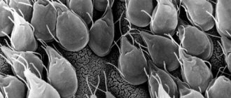
Opisthorchid in fish is a common occurrence today. It is almost impossible to determine the infection externally. River fish, especially the carp family, are susceptible to the disease. Infection occurs as a result of eating raw fish or improperly preparing it. For a long time the disease proceeds hidden. Affects the liver, pancreas, and gall bladder. Causes cancer. Characterized by frequent exacerbation.
Pathogenesis of opisthorchiasis
Pathological processes in opisthorchiasis affect many organs and systems.
Allergy
Opisthorchid waste products (toxins) in the host’s body cause sensitization (hypersensitivity) and the development of an allergic reaction. Acting as antigens, they interact with cells of the host's immune system, T-lymphocytes and macrophages, which triggers complex processes leading to an increase in the level of immunoglobulins E (Ig E). Biologically active substances are released from mast cells, which rush to the foci of immune inflammation, as a result of which proliferative-exudative reactions develop in the skin, mucous membranes, joints, lungs, bronchi, organs of the digestive system, heart and other organs.
The permeability of blood vessels in the liver and pancreas increases. The vascular wall, infiltrated with eosinophils, swells. Foci of necrosis appear in the organs. Antigenic pancreatitis and hepatitis develop. Hyperplasia of RES cells leads to enlargement of lymph nodes.
Focal and diffuse changes of an allergic nature appear in the stomach and intestines, and massive eosinophilic infiltrates appear.
Areas of allergic dermatitis appear on the skin.
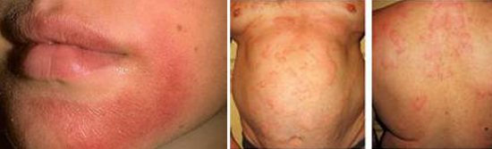
Rice. 3. In the acute stage of opisthorchiasis, the body’s allergic reaction comes to the fore. The photo shows signs of opisthorchiasis - rashes on the skin of an allergic nature.
Mechanical impact
At the stage of sexual maturity, the cat fluke parasitizes the gall bladder, bile ducts and pancreatic ducts. The parasite has 2 suckers that help it stay in the host’s body - oral and abdominal. The oral sucker and cuticular spines are located at the head end. When fixed, the suction cups cause mechanical damage to the mucous membrane - they retract and pinch it. Young individuals are fixed with the help of cuticular spines, which also leads to mechanical damage to the walls of the gallbladder, bile ducts and pancreatic ducts and disruption of their blood supply. Multiple bleeding erosions appear on the walls of the ducts. Stimulation of regenerative-hyperplastic processes leads to their thickening and the development of connective tissue in the organ itself.
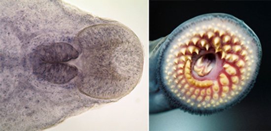
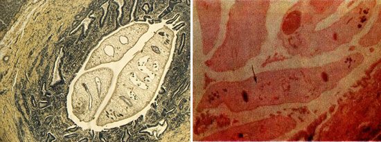
Rice. 4. Longitudinal section of the oral sucker and pharynx (photo on the left) and the oral sucker of opisthorch with cuticular spines (photo on the right).
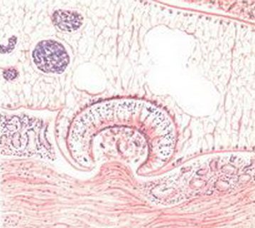
Rice. 5. Opisthorchiasis of the liver. The parasite draws in the wall of the bile duct (microscopic specimen).
Obturation
Opisthorchids do not reproduce in the host's body, but with constant eating of infected fish, the parasites accumulate and lay eggs. One parasite lays up to 900 eggs per day. Their accumulation together with the desquamated epithelium leads to obstruction of the passages, as a result of which the outflow of bile and pancreatic juices is hampered. In the chronic course of the disease, the ducts degenerate cystically. Their walls thicken, and connective tissue develops around them. A secondary infection often occurs. Damage to the bile ducts sometimes leads to the development of cholangitis cirrhosis of the liver.
Toxic-allergic reactions and mechanical effects of helminths are the main links in the pathogenesis of opisthorchiasis.
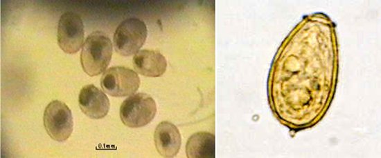
Rice. 6. Opisthorchid eggs.
Secondary infection
Stagnation of bile contributes to the colonization of the bile ducts by bacteria. Bacteria enter the bile ducts along with parasites from the intestines, and are also introduced hematogenously and cause the development of bacterial cholecystitis, inflammation of the bile ducts (cholangitis), chronic hepatitis, and rarely, cholangiolytic cirrhosis.
Oncology
Glandular hyperplasia, which developed as a result of prolonged exposure to opisthorchids on the walls of the bile ducts, creates the basis for the development of cholangiocarcinoma. Opisthorchiasis, caused by Opisthorchis viverrini, is included in the list of category 1 carcinogens.
In the acute stage of opisthorchiasis, the allergic reaction of the body, which has developed to the waste products of parasites, comes to the fore; in the chronic stage, the long-term mechanical impact of mature parasites comes to the fore.
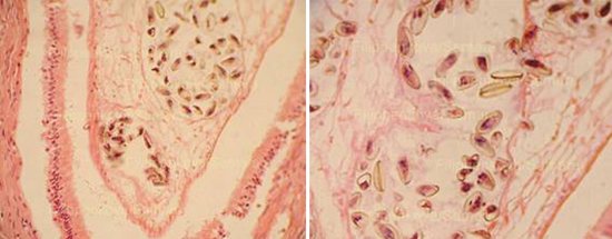
Rice. 7. The photo shows fragments of the uterus of a Siberian fluke in the bile ducts with a mass of eggs (microscopic specimen).
Incubation period
Opisthorchid enters the human or carnivorous body by consuming thermally untreated or insufficiently processed fish of the carp family in the form of metacercariae enclosed in a shell - a cyst. In the gastrointestinal tract, the membrane of the metacercariae dissolves and the parasites penetrate the biliary tract, gall bladder and pancreatic ducts, where within 10 - 12 days they develop to a sexually mature state and begin to lay eggs. The incubation period for opisthorchiasis ranges from 2 to 4 weeks (5 to 42 days). It is believed that on average 21 days pass from the moment of infection to the appearance of the first signs and symptoms of the disease.
What is opisthorchiasis
A disease associated with infection with roundworms - worms. Foci of varying degrees of intensity are due to several factors:
- the presence of bodies of water in a certain area;
- pollution of water sources with sewage;
- habits of the population to consume raw fish, lightly salted.
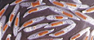
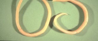
The causative agents of opisthorchiasis are considered to be 2 species of annelids - Opisthorchis felineus, O. viverrini. The first type of worms is the most dangerous for humans; it also provokes the disease. An adult reaches a size of 18 mm. The body is about 1.5 mm wide. The parasite reproduces up to 900 eggs per day. The life cycle of worms is quite complex. Provides 1 definitive host and two intermediate hosts. This explains their wide distribution in nature. The original host is a mollusk. Worms develop in it for about a year. Opisthorchis tends to infect shellfish that are found in stagnant water or with a slow current. A certain part of them die in the water. The intermediate host is fish. The question arises, what kind of fish do opisthorchids live in? They can infect:
- roach;
- ide;
- bream;
- subdust;
- tench;
- verkhovka;
- dace;
- rudd;
- chub;
- blue;
- uklel;
- carp;
- white-eye;
- silver bream;
- saberfish;
- spike.
Infected fish show no obvious symptoms of infection. Which makes it especially dangerous. The final owner is a person. The parasite most often settles in the liver, the gallbladder is in second place, and a certain proportion falls on the pancreas.
Signs and symptoms of opisthorchiasis in the acute stage
In the acute stage of opisthorchiasis, the allergic reaction of the body, which has developed to the waste products of parasites, comes to the fore, manifested by granulomatous hepatitis, pancreatitis, pneumonitis of an antigenic nature and eosinophilic infiltrates in a number of other organs and tissues. The acute stage of the disease with mild to moderate severity lasts 1 - 2 weeks, with severe disease - 4 - 8 weeks or more.
Clear signs and symptoms of opisthorchiasis appear when a person enters the area where the disease is spreading for the first time. Obliterated signs and symptoms of opisthorchiasis are observed in residents permanently residing in the parasitosis zone.
- The acute stage of the disease begins with an increase in body temperature.
- Urticaria-type rashes are observed on the skin.
- Muscle-joint pain appears.
- After some time, the patient begins to experience pain in the right hypochondrium and at the point of the gallbladder, and the liver enlarges. Over time, antigenic pancreatitis and hepatitis develop, and the lymph nodes enlarge.
- Symptoms of damage to the gastrointestinal tract appear: nausea, heartburn, pain in the stomach, stools become frequent and loose, flatulence is noted, and appetite decreases. Erosive gastroduodenitis develops.
- Some patients develop symptoms of damage to the bronchopulmonary system of an allergic nature - antigenic pneumonitis and asthmatic bronchitis.
Signs and symptoms of mild opisthorchiasis
With a mild degree of the disease, after a sudden increase to 380C, the body temperature drops to low-grade and lasts for 1 - 2 weeks. Increased fatigue, weakness, pain in the right hypochondrium, vague abdominal pain and loose stools are the main symptoms of mild opisthorchiasis.
Signs and symptoms of moderate opisthorchiasis
With opisthorchiasis of moderate severity, after a sudden increase to 380C, the body temperature drops to subfebrile and lasts for 2 - 3 weeks. The above-described symptoms are accompanied by muscle and joint pain, allergic rashes appear on the skin, vomiting and diarrhea, signs of bronchial damage - bronchitis with an asthmatic component, the spleen, liver and lymph nodes become enlarged.
Signs and symptoms of severe opisthorchiasis
In severe cases of opisthorchiasis, the patient experiences pronounced symptoms of intoxication, body temperature reaches 39.0 - 39.5 ° C, urticaria-type skin rashes are abundant, some patients develop Quincke's edema, the patient is inhibited or excited, the liver and spleen are enlarged, Liver tests increase, jaundice appears, pain in the right hypochondrium is persistent and paroxysmal, lymph nodes enlarge, abdominal bloating, nausea, vomiting and diarrhea are noted, symptoms of damage to the bronchi and lung tissue appear - cough, attacks of suffocation, shortness of breath and chest pain.
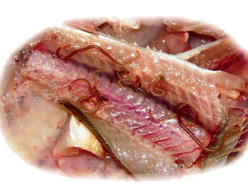
Rice. 8. The photo shows three opisthorchids in the human pancreatic duct.
Variants of the course of opisthorchiasis
Opisthorchiasis, which occurs in severe form, can have several variants of the course.
Typhoid-like variant
The typhus-like variant most clearly reflects the allergic basis of the disease. Acute onset, high body temperature, chills, swollen lymph nodes, muscle and joint pain, heart pain, nausea and vomiting, cough and skin rashes are the main symptoms of opisthorchiasis of the typhoid-like course. The disease lasts up to 2 - 2.5 weeks.
Hepatocholangitic variant
Against the background of high body temperature, the patient develops symptoms of liver damage: pain in the right hypochondrium, nausea, vomiting. The liver enlarges. Liver tests increase. Icterus (yellowness) of the sclera is noted.
Gastroenterocolitic variant
The disease occurs with symptoms of gastritis, colitis and stomach ulcers. Decreased appetite, nausea, rarely vomiting, abdominal pain of undetermined localization and loose stools are symptoms of opisthorchiasis of the gastroenterocolitic variant.
Variant of the course of opisthorchiasis with damage to the respiratory tract
A variant of the course of opisthorchiasis with damage to the respiratory tract is registered in every third patient. Mucous discharge from the nose appears, swelling and redness of the back wall of the pharynx is noted, symptoms of damage to the bronchi and lungs appear: cough, asthma attacks, shortness of breath, chest pain, and less often - symptoms of bronchial asthma.
Asymptomatic course of opisthorchiasis
Sometimes at an early stage opisthorchiasis is asymptomatic. However, there is always an increase in the number of eosinophils in the blood.
Due to the development of immunological resistance in permanent residents of the focus of opisthorchiasis, the acute phase of the disease may be absent, or it may be much milder than among visitors.
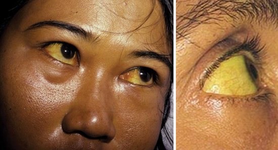
Rice. 9. In severe cases of opisthorchiasis, liver function is disrupted, and yellowness of the sclera and skin appears.
Prevention measures
Opisthorchiasis metacercariae survive well at both low and high temperatures. For example, when fish is frozen to a temperature of -13°C, the death of the larvae occurs only after a month. If you bring the freezing to lower temperatures, for example to -30°C, then after 6 hours the larvae will die, and at a temperature of -40°C it will take 3 hours to eliminate them.
From this we can conclude that with simple heat treatment inside the fish, complete death of the parasites cannot occur. At a cooking water temperature of +40°C, only part of the larvae are destroyed. It is necessary to heat the water to +100°C, then after 15 minutes all the parasite larvae will die. But if you fry the fish, it will take 20 minutes for the worms to die. It is advisable to cover the pan with a lid, this will more guarantee their complete destruction.
The safest method of preparing seafood is hot smoking technology. If this is cold smoking, then the fish is pre-frozen or salted for 2 weeks (20 kg of salt is taken per 100 kg of product).
Chronic opisthorchiasis
If in the acute stage of the disease the allergic reaction of the body, which has developed to the waste products of parasites, comes to the fore, then in chronic opisthorchiasis the pathogenesis is based on the long-term mechanical impact of mature parasites.
- Attachment of opisthorchid to the walls of the bile and pancreatic ducts and the walls of the gallbladder leads to a violation of their integrity, the appearance of many bleeding erosions, and the development of inflammation. Stimulation of regeneration processes leads to expansion of the ducts and thickening of their walls; areas of necrosis and sclerosis appear in the parenchyma of organs.
- Accumulations of parasite eggs, desquamated epithelium and mucus impede the outflow (promotion) of bile and pancreatic juices through the ducts, which leads to their expansion and the development of cholangio- and canaliculoectasis.
Chronic opisthorchiasis lasts a long time - 15 - 25 years or more. Periods of remission are followed by periods of exacerbation. The disease in chronic form occurs mainly among residents of regions where helminthiasis is most common. In 100% of cases, opisthorchiasis is localized in the bile ducts, in 60% of cases - in the gallbladder, in 36% of cases - in the pancreatic ducts.
In an uncomplicated course, chronic opisthorchiasis has a benign course. Liver function is slightly impaired. Basically, the pathological process is localized in the biliary tract. Often chronic opisthorchiasis is combined with chronic pancreatitis, gastritis, gastroduodenitis. In some patients, the disease is mainly manifested by symptoms of cholecystitis and cholangitis, in others the disease occurs with symptoms of enzyme deficiency, in others - with symptoms of allergies and toxicosis. Sometimes there is an asymptomatic course of chronic opisthorchiasis.
After treatment of chronic opisthorchiasis, irreversible changes remain in the liver and biliary tract, pancreas and its ducts, stomach and other organs. Health measures aimed at improving digestion processes in this case become of great importance.
Signs and symptoms of opisthorchiasis with liver damage
In chronic opisthorchiasis, the liver is rarely affected. Sometimes small focal lesions are recorded. The liver is often of normal size or slightly enlarged and hardened. In severe cases, liver function is significantly disrupted, patients develop yellowness of the skin and mucous membranes, and biochemical parameters increase - bilirubin, transaminases, etc.
The course of opisthorchiasis is aggravated by the presence of viral hepatitis, other infectious diseases and exposure to toxic factors.
With a long course of helminthiasis, in some cases chronic hepatitis, liver cirrhosis and hepatocellular carcinoma develop.
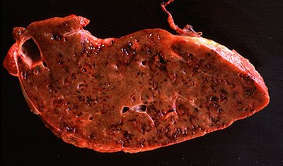
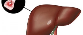
Rice. 10. Liver damage due to opisthorchiasis.
Signs and symptoms of opisthorchiasis with damage to the gallbladder and bile ducts
With opisthorchiasis, the gallbladder and bile ducts are primarily affected. Biliary dyskinesia, cholangitis and cholecystitis develop. The patient develops pain in the right hypochondrium and attacks of biliary colic. The gallbladder increases in size. The duodenal contents contain a lot of mucus, epithelial cells, leukocytes and parasite eggs. Biliary peritonitis and abscess are severe complications of the disease.
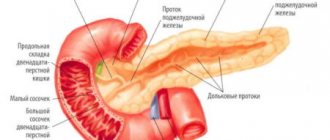
Signs and symptoms of opisthorchiasis with damage to the pancreas
In chronic opisthorchiasis, in 36% of cases, the pancreatic ducts and the organ itself are affected. Glandular hyperplasia of the epithelium develops in the walls of the ducts, the walls themselves thicken, and areas of sclerosis and necrosis appear in the pancreas. Severe complications of the disease are destructive pancreatitis and pancreatic cancer.
When the ducts and the pancreas itself are damaged, girdling pain and nausea occur.
Signs and symptoms of opisthorchiasis in the gastrointestinal tract
With opisthorchiasis, the gastrointestinal tract is often involved in the pathological process. Nausea, heartburn and pain in the stomach appear, stools become frequent and liquid, flatulence is noted, appetite decreases, erosive gastroduodenitis develops, and the acidity of gastric juice decreases. A high content of lactic acid in feces leads to itching in the anus. Ahilia creates conditions for the development of secondary flora.
Signs and symptoms of opisthorchiasis with damage to the central nervous system
With chronic opisthorchiasis, in some cases, astheno-neurotic syndrome develops. The patient experiences weakness, dizziness, sleep disturbances, mental and physical fatigue, trembling of fingers, tongue and eyelids, irritability, emotional instability, frequent mood swings and depression. In the case when astheno-neurotic syndrome comes to the fore, the patient is diagnosed with “vegetative neurosis” or “neurocirculatory dystonia”.
Signs and symptoms of opisthorchiasis with the development of allergic syndrome
In some patients with chronic opisthorchiasis, due to the individual characteristics of the immune system, an allergic syndrome may come to the fore. Opisthorchid waste products (toxins) act as antigens. They interact with cells of the host immune system, T lymphocytes and macrophages, which triggers complex processes leading to increased levels of immunoglobulins E (Ig E). Biologically active substances are released from mast cells, which rush to the foci of immune inflammation, as a result of which proliferative-exudative reactions develop in the skin, mucous membranes, joints, lungs, bronchi, organs of the digestive system, heart and other organs.
The cat fluke lives in the human body from 10 to 20 and even up to 40 years, in the cat’s body - 3 or more years.
Rice. 11. The photo shows opisthorchid in the bile duct of a human liver.
Infection with opisthorchiasis from fish
Formed individuals produce eggs in huge quantities. They, together with the feces of their owners, end up in the external environment, a body of water. The eggs float in the pond and are sucked up by mollusks. For a certain period of time they develop in the body of the mollusk. Then they enter the water again. Larvae with a tail are well retained in standing water or with a weak current. When they see a fish, they penetrate its body, possibly penetrating through the gills. In fish, the larvae already live without a tail. Concentrated in the muscles. Infection with opisthorchiasis in humans occurs only through food. Raw meat that is not properly cooked. Becomes a source of infection.
The larvae go through the final stage of development. This will take from 14 to 21 days. After which the disease makes itself felt. Its symptoms are similar to most diseases of the digestive tract. A person lives most of his life with adult worms inside, treats gastrointestinal diseases, but the therapy does not give the desired result.
The larva in the human duodenum, under the influence of gastric juice, is freed from its previous covering. Gradually it makes its way to the liver, gall bladder, and some of it enters the pancreas. Infection with opisthorchiasis occurs within 5 hours. For another 14 days the larva develops and becomes a sexually mature individual. The disease continues for years. In the absence of proper treatment - up to 30 years.