The liver is one of the most important organs in the human body, as it performs many functions that ensure normal life. Therefore, a tumor of this organ is always characterized by a severe course and high mortality. However, the disease occurs quite rarely, mainly at 4 stages of the oncological process of another localization. Primary adenocarcinoma of the liver tissue occurs no more often than 25% of cases
. Secondary lesions penetrate the liver through lymphogenous, hematogenous, retrograde routes, or through direct tumor growth into the organ tissue. Metastases in the liver most often give rise to the following adenocarcinomas:
- stomach wall cancer;
- lung tumor;
- damage to the rectum and large intestine;
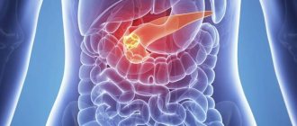
- tumor neoplasms of the pancreas;
- breast cancer;
- foci of malignancy in the kidneys.
Liver cancer is also characterized by a reverse spread. That is, those organs that metastasize to a given organ are primarily affected by tumor cells if adenocarcinoma initially occurs in the liver.
Reasons for the development of adenocarcinoma in the liver
Reliable reasons for the formation of tumor tissue in any human organ have not yet been identified. The following predisposing factors are characteristic of adenocarcinoma in the liver:
- chronic viral hepatitis (diseases gradually change the structure of the cellular structure of hepatocytes, which leads to malignancy);
- cirrhosis or gradual destruction of organ cells (mass death of hepatocytes significantly increases the risk of adenocarcinoma);
- alcoholism (with metastases of adenocarcinoma in the liver, this factor can aggravate the rate and severity of cancer);
- syphilitic damage to the body and diabetes mellitus;
- cholelithiasis (stagnation of bile and inflammatory processes in the liver tissues provoke malignant degeneration);
- parasitic and fungal infections;
- the presence of liver adenocarcinoma in close relatives.
Additional risk factors include various chemical compounds and negative physical effects (ionizing radiation), which are quite rare.
Classification according to forms and degrees of differentiation
Liver cancer can have three forms:
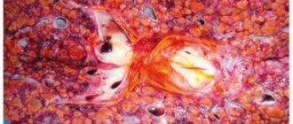
- Nodal. This adenocarcinoma occurs in 65% of cases and is characterized by the appearance of nodules delimited from healthy tissue. There may be several neoplasms, and they contribute to the enlargement of the organ.
- Massive. In morphology it does not differ from the nodular variant, but reaches large sizes and most often represents a single node. The typical location is the right lobe of the liver.
- Diffuse. A rare type of adenocarcinoma in which cells replace healthy hepatocytes. The organ does not enlarge.
The degree of differentiation of cellular structures is important, of which there may be three or four types (some sources define the poorly differentiated and undifferentiated variants as one group). The classification includes the following forms of adenocarcinoma:
- Highly differentiated
. Most often it is a primary cancer, the cells of which are similar to healthy liver tissue. - Moderately differentiated
. A transitional option, in which differences from healthy tissue are noticeable, but not sufficiently pronounced. - Poorly differentiated
. The development of poorly differentiated liver adenocarcinoma is caused by metastasis of cells from the primary lesion. Tumor tissue differs sharply from healthy tissue and has practically no structures similar to it. - Undifferentiated
(conditionally distinguished from the previous type). This type differs from a poorly differentiated tumor only in that histologists cannot determine the original origin of the malignant tissue.
Clinical picture of adenocarcinoma
With secondary damage to the liver tissue by tumor cells, the disease is not diagnosed immediately, but with primary malignancy, the latent period is additionally masked by the clinic of the underlying pathology, for example, cirrhosis.
When adenocarcinoma of any localization appears, nonspecific symptoms of cancer are primarily formed: loss of appetite and rapid weight loss, decreased performance and apathy, low-grade fever of a chronic nature, dyspeptic syndrome for no apparent reason. The main, but not specific enough sign of liver adenocarcinoma is yellowness of the skin and mucous membranes with pain in the right hypochondrium, which gradually worsens. Patients' urine becomes darker and their stool becomes lighter. Regional lymph nodes enlarge and a pathological change in the size of the liver is noted. In addition, petechiae appear on the skin of patients with adenocarcinoma, and fluid begins to accumulate in the abdominal cavity (ascites is formed). The clinical picture of the disease corresponds to cirrhosis, that is, the breakdown of liver tissue, or severe acute hepatitis. Only diagnostic measures help to suspect cancer.
Clinical picture of the disease
The disease develops slowly and does not have specific symptoms for a long time. The composition of the neoplasm resembles healthy tissue. A highly differentiated tumor makes itself felt as it grows and puts pressure on the nerve endings. Symptoms depend on the location of the outbreak:
- An enlarged node in the pancreas is manifested by weight loss. The patient experiences a feeling of overeating, abdominal pain, and notices abnormal bowel movements. The skin acquires a yellow tint.
- A highly differentiated tumor in the stomach is characterized by symptoms of poisoning (nausea, vomiting, diarrhea), bloating, lack of appetite, a feeling of heaviness and pain in the epigastric region. If the node reaches a large volume and puts pressure on the wall of the organ, perforation followed by peritonitis is possible. The patient will feel acute pain, and the body temperature will rise sharply.
- If the colon is damaged, there is aching pain, stool with mucus, pus and streaks of blood, lack of appetite, sudden weight loss, lethargy and high body temperature. With a node with ulceration, an inflammatory process develops.
- Typically, a neoplasm in the rectum occurs in representatives of the stronger sex. Localized near the anus. The patient also experiences pain during bowel movements, mucus and pus are released, flatulence is noted, and streaks of blood are present in the stool. At an early stage of oncology, the symptoms are similar to hemorrhoids.
Adenocarcinoma tumor in the rectum
- With pathology of the sigmoid colon, symptoms are characteristic of diseases of the large intestine.
- Tumors of the sigmoid colon provoke rumbling in the abdomen, cramping pain, partial bloating, abnormal stool, and bleeding.
- Uterine tumors are characterized by prolonged bleeding of various origins. The woman notices heavy discharge that differs from normal in color and smell.
- If the prostate is damaged, pain occurs when urinating, there is no erection, and the bladder feels constantly full.
- Lung adenocarcinoma is expressed by cough with mucus, shortness of breath and chest pain.
- Tumors with ulceration allow pathogenic organisms to penetrate the organ walls. Because of this, infectious diseases develop.
- Neoplasms with decay are characterized by arrhythmia, bradycardia, frequent fainting, acute renal failure, dyspepsia, nausea and vomiting.
Well-differentiated adenocarcinoma is characterized by general symptoms:
- Exhaustion of the body in a short period of time.
- An increase in body temperature against the background of good health.
- Pain in the affected organ.
- Lethargy and unreasonable fatigue.
- Decreased brain activity, dizziness.
Stages of cancer
According to the international classification of diseases, there are 4 stages of any cancer, each of which has its own characteristics:
- First
. Characterized by sizes up to 2 cm, therefore it does not manifest itself clinically. The tumor does not affect the functioning of the liver and does not affect neighboring structures, and therefore is not diagnosed. - Second
. The size of the cancer still does not exceed 2 cm, but damage to adjacent vessels may occur. A variant of the second stage of adenocarcinoma is the presence of several foci without the involvement of vascular structures. - Third
. The cancer grows more than 2 cm but is limited to one lobe of the liver. There may be several foci, the vessels are not always affected, but the lymph nodes become enlarged. The tumor can be felt on the anterior abdominal wall. - Fourth
. Stage 4 of liver adenocarcinoma is critical, which is characterized by the presence of distant metastases and severe damage to both lobes of the organ.
Establishing the stage of the oncological process determines the calculation of the prognosis and survival of the patient, subject to proper treatment. The last stage is an indication for palliative care with symptom relief.
Etiology of the disease
The exact cause of the disease is being determined. Scientists believe that cancer develops against the background of certain factors:
- It is most likely that the malignant process is caused by disturbances in the human genetic code, which means that the possibility of developing cancer begins in the prenatal period of development.
- Hereditary predisposition.
- Chronic inflammatory diseases of internal organs.
- Chronic disorders in the functioning of organs.
- Overeating or undereating.
- Bad eating habits, for example, smoked foods, fried fatty meats, foods that contain preservatives and chemical additives.
- Alcohol and nicotine addiction.
- Work in metallurgical and coal production, where a person constantly inhales heavy components.
- Prolonged contact with radiation and ultraviolet radiation.
- Viral diseases, especially HPV.
Degrees of tumor differentiation
The course of the disease depends on the degree of malignancy of the cancer:
- A well-differentiated (G1) tumor contains atypical cells that differ slightly from normal ones. The shape of the core is different. The disease develops over a long time and does not provoke the growth of metastases. Treatment is effective at any stage of development.
- The moderately differentiated (G2) form of cancer contains a larger number of cells that differ from normal in size and shape. A more aggressive course is observed. Relapse may occur after treatment.
- Metastatic adenocarcinoma is a poorly differentiated (G3) node consisting entirely of abnormal cells. Due to rapid division, cells disperse through the bloodstream and lymph to all parts of the body. Metastasis begins in the early stages of development. Treatment does not bring maximum results. The prognosis for life is negative. In this regard, the patient’s life expectancy is reduced to a year.
Stages of cancer development
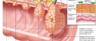
Adenocarcinoma develops during 4 stages:
- Stage 1 is characterized by the formation of a node up to 2 cm in size. The epithelial layer of the organ is affected, the tumor does not extend beyond its limits. There are no symptoms or metastases.
- At stage 2, the size of the lesion reaches 5 cm. It does not extend beyond the organ, but cancer cells can spread to regional lymph nodes.
- At stage 3, regional lymph nodes become enlarged. The tumor grows into the walls of the organ. Metastases do not spread.
- At the terminal stage of cancer, the entire body is involved in the process. Secondary lesions spread throughout the body. The patient can die at any time.
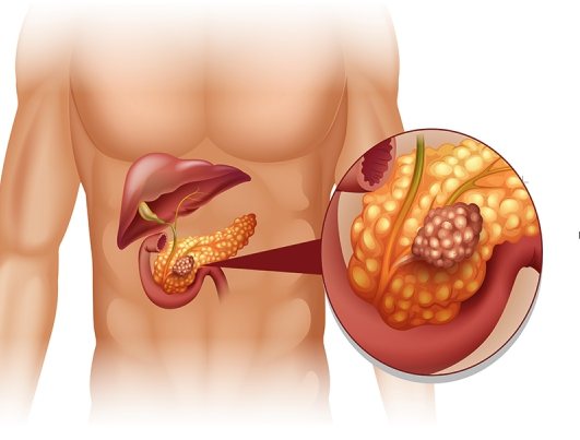
Pancreatic adenocarcinoma
Types of tumors depending on the primary location of cancer cells:
- Mucinous tumor usually affects the endometrial wall. The cystic cells of the neoplasm produce mucus. Mucins quickly spread to neighboring organs.
- The neoplasm of a glandular solid structure is represented by trabeculae with layers of connective tissue.
- A tubular type neoplasm consists of tubular cells.
- A scirrhous tumor grows from the stroma.
- Papillary papillary tumor usually affects tissue of the thyroid gland, ovary and kidney.
- The prostate gland is characterized by an acinar form of cancer. The node produces fluid, which, spreading through the tissues, infects neighboring organs with cancer.
- Clear cell pathology develops in the genitourinary system in the form of polyps. Contains various forms of cells, for example, solid, papillary, tubular and cystic.
- Colon cancer is an invasive tumor that damages all parts of the organ and extends beyond its boundaries.
- The neoplasm in the esophagus consists of epithelial cells. As a rule, it develops in men against the background of bad habits.
- Adenocarcinoma of the mammary glands shows an infiltrative nature.
Principles of diagnosing adenocarcinoma
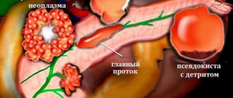
Adenocarcinoma with liver metastases is difficult to detect and requires an integrated approach to diagnosis. First, non-specific studies are carried out. A biochemical blood test will show an increase in enzymes and bilirubin, which indicate organ damage. An ultrasound will help visualize the tumor
. A specific test for adenocarcinoma of the liver localization is the detection of alpha-fetoprotein (tumor marker). Its presence indicates the formation of a primary focus in this organ. A computed tomography scan is also performed, without which a diagnosis according to the TMN classification is impossible. Histology remains a mandatory research method, for which tumor tissue taken during laparoscopy or targeted biopsy is sent.
Liver cancer is characterized by late diagnosis, which is due to:
- long latent period of the disease;
- masking the clinical picture as background pathologies;
- slow progression of the tumor (in the case of primary malignancy).

Diagnosis of moderately differentiated adenocarcinoma
To determine the type, location, stage of development, and degree of malignancy of the tumor, oncologists prescribe a number of laboratory and instrumental studies:
- General analysis of urine and blood. The results show the presence of an inflammatory process in the body. If an oncological process is suspected, the ESR indicator is important.
- A biochemical blood test determines the performance of the kidneys, liver, gallbladder, pancreas and other organs.
- For neoplasms in the genital organs, the patient donates blood to determine hormone levels.
- Tests for tumor markers are informative, but show the disease at later stages of development.
- To identify tumors of the uterus, ovaries, prostate and rectum, a manual examination is performed.
- Ultrasound examination detects changes in the pelvic organs, gastrointestinal tract and lymph nodes.
- X-rays are used to diagnose cancer of the lungs and other organs. When examining the gastrointestinal tract, the patient is injected with a contrast agent - barium.
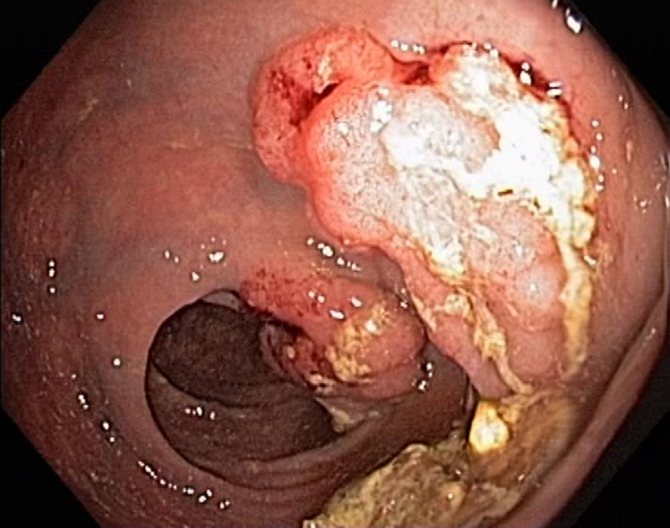
- Endoscopic examinations allow you to examine organs from the inside and take a biopsy without damaging the skin. Relevant for diagnosing the stomach, intestines, bladder and respiratory system.
- Laparoscopy is a minimally invasive diagnostic procedure. A laparoscope with a camera and a flashlight at the end is inserted into the body through a puncture in the skin. The doctor gains access to the internal organs. The method is used to remove small tumors.
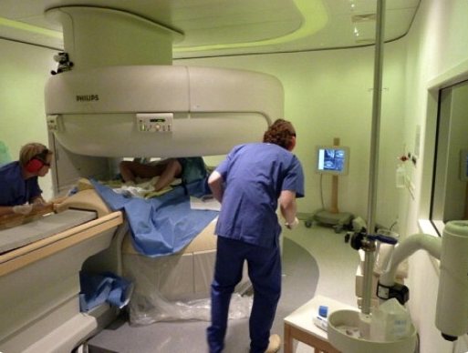
- Magnetic resonance and computed tomography perform layer-by-layer scanning of human organs and systems. Tumors are detected along with the method of blood supply, changes in organs, metastases in lymph nodes and bone tissue.
- Angiography evaluates the condition of blood vessels.
- Histological examination is final for determining the diagnosis. Microscopy of a biopsy specimen determines the nature of the neoplasm, the degree of differentiation, how damaged the normal tissue cell is and the stage of the process.
Liver cancer treatment
Oncologists have developed three main methods for treating liver adenocarcinoma: surgery (tumor removal), radiation, and chemotherapy. Most neoplasms in the liver tissue remain operable and therefore must be removed. Resection of the organ is performed with excision of the affected area and nearby lymph nodes. Patients who have fluid accumulation in the abdominal cavity, cirrhosis or vascular thrombosis are considered inoperable. Such patients are given symptomatic treatment, which only slightly stops the progression of the disease. Liver transplantation is considered a fairly successful treatment method. However, it is completely ineffective for patients with secondary lesions. In addition, an organ transplant does not guarantee that adenocarcinoma will not recur
.
Treatment with medications or radiation does not bring the expected positive effect, but can be used to relieve symptoms, affect metastases and stop cancer growth.
These treatments are used in patients with no chance of recovery and in those on the transplant waiting list. In the latter case, conservative therapy reduces the risk of developing relapses in the future, as it helps to destroy tumor cells circulating in the blood.
Free quota surgery: how to get it and what’s changing in 2020
Cancer treatment >> Types of cancer >> Liver adenocarcinoma is a malignant neoplasm that is formed from the glandular tissue of the liver or, which is much more common, appears as a result of metastasis of a tumor with primary localization in another organ (stomach, uterus, ovaries, colon, lungs, mammary gland).
Kinds
Depending on the level of tumor differentiation, three types of adenocarcinoma are distinguished:
- highly differentiated - neoplasm cells are practically unchanged compared to normal tissue cells; this disease has the most benign course; - moderately differentiated - occupies an intermediate position; - poorly differentiated - the tumor has maximum malignancy, metastasizes early and is difficult to treat.
Risk factors
- male gender - this disease occurs 3 times more often in men than in women; - smoking; — chronic viral hepatitis; - cirrhosis of the liver; - steatohepatitis; - contact with toxic substances, especially aflatoxins and arsenic.
Symptoms
The clinical picture of liver adenocarcinoma contains both general signs of malignant neoplasms and manifestations characteristic only of this type of cancer. These include: loss of appetite, nausea, vomiting, progressive weakness, anemia, a feeling of heaviness and pain in the epigastrium or right hypochondrium, enlarged liver or a palpable node in the right hypochondrium, obstructive jaundice, frequent nosebleeds, an unreasonable increase in body temperature up to 37C, ascites.
Metastases
Liver adenocarcinoma most often metastasizes to the liver itself, nearby lymph nodes, peritoneum, pancreas, kidneys, lungs, pleura, and bones.
Diagnosis and treatment
To diagnose liver adenocarcinoma, the following examination methods are used: blood serum analysis for alpha-fetoprotein content, liver ultrasound, and MRI, positron emission tomography, angiography, as well as liver biopsy followed by histological examination of the biopsy.
Treatment of liver adenocarcinoma is carried out using various methods, usually used in combination. The most radical method is liver transplantation; partial liver resection is also possible, performed in approximately 10% of patients with advanced stages of the disease. If liver resection is not feasible while the patient is on the transplant waiting list, percutaneous ethanol injection or radiofrequency ablation techniques are used to delay tumor growth.
If transplantation and surgical treatment are impossible, patients with liver adenocarcinoma are prescribed chemoembolization (regional chemotherapy) with blocking the blood flow of the blood vessel feeding the tumor and simultaneous administration of a chemotherapy drug. Systemic chemotherapy and radiation therapy for this diagnosis do not have significant effectiveness and are used in combination treatment.
Forecast
Liver adenocarcinoma has a generally poor prognosis. The five-year survival rate for patients with this diagnosis averages 10%; if the disease is detected at the first stage and treatment is started in a timely manner, this figure reaches 40%.
Prognosis for adenocarcinoma in the liver
Regardless of the timeliness of diagnosis or the adequacy of treatment, the prognosis for liver adenocarcinoma remains unfavorable.
The defeat of this organ is characterized by many dysfunctions, many of which cannot be restored. Even full treatment and compliance with all doctor’s recommendations for adenocarcinoma in the first stage determines only a 40% chance of five-year survival. Later diagnosis raises the mortality rate to almost 100%. Found a mistake? Select it and press Ctrl + Enter