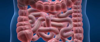
general description
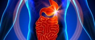
Speaking about what gastric adenocarcinoma is, it should be noted: this is a form of cancer classified not by clinical manifestations, but based on which cells have changed under the influence of oncogenic factors, giving rise to the tumor. To do this, it is studied under a microscope, conducting a histological examination. Adenocarcinoma is a derivative of glandular epithelium. According to the WHO histological classification, there are:
- Tubular adenocarcinoma of the stomach, which can be highly differentiated (that is, the cells do not differ much from normal glandular tissue) and moderately differentiated (the cells are greatly changed, but you can “guess” their origin).
- Poorly differentiated gastric adenocarcinoma. Differentiation is the process of cell maturation, during which they acquire a structure and properties characteristic only of a certain tissue. In poorly differentiated carcinoma, they are so “immature” that they have practically nothing in common with normal glandular epithelium.
The lower the cell differentiation, the more malignant and aggressive the tumor behaves; therefore, other things being equal, well-differentiated gastric adenocarcinoma has a more favorable prognosis than poorly differentiated one.

The so-called mucinous adenocarcinoma of the stomach or “mucosal cancer” is distinguished separately, which is characterized by increased mucus formation and early metastasis.
Diagnostics
The main goal of diagnosis is to identify the degree of differentiation and localization of the tumor. After conducting a personal examination and clarifying the medical history, the gastroenterologist or oncologist will refer you for the following types of studies:
- A complete blood count can detect anemia.
- Testing for tumor markers helps determine the severity of gastric damage.
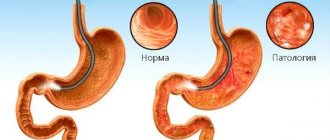
- Gastroscopy visualizes the pathological focus, and a biopsy will allow cytological and histological studies of the tissue sample taken.
- X-ray with a contrast agent will make it possible to assess the extent of damage to the stomach wall.
- Computed tomography (CT) will determine the presence of metastases in distant organs and lymph nodes.
- Endoscopic ultrasound will help determine the extent of tumor penetration into the organ wall, lymph nodes, and nearby tissues.

Causes
Cancer is cells whose genetic structure has been altered either due to a hereditary defect or under the influence of external factors called carcinogens. Under the influence of these factors, cells lose the ability to naturally die (apoptosis) and differentiate (maturation), but acquire the ability to multiply uncontrollably. In the case of gastric adenocarcinoma, the main carcinogenic factor is considered to be Helicobacter Pilory infection. This bacterium is stable in an acidic environment and causes long-term inflammation of the mucous membrane - gastritis, which over time turns into atrophic, and then into cancer.
Another predisposing factor is dietary habits: addiction to spicy, salty, pickled foods, sausages and canned food, the preparation of which uses nitrites (which turn into nitrates in the stomach). The role of dietary habits in the development of cancer can be judged by the fact that among the second generation of emigrants from Japan, the incidence of stomach cancer decreases significantly - despite the fact that this country has retained the first place in incidence in the world for many years.
Precancerous conditions also include:
- Ménétrier's disease is a rare form of hyperplastic gastritis;
- adenomatous polyps - damage to the gastrointestinal tract, often of a hereditary nature;
- gastric resection - according to statistics, 25 years after surgery, the likelihood of developing adenocarcinoma increases 8 times.
On average, at least 10 years pass from the beginning of the growth of a malignant tumor to the appearance of the first clinical signs.
Treatment of well-differentiated gastric adenocarcinoma
The choice of treatment for well-differentiated adenocarcinoma depends on the location, size and histological component of the tumor. The main method is surgical excision of the tumor. Removal of a highly differentiated tumor is carried out together with part of the healthy tissue.
At later stages, oncologists prescribe subtotal or total resection. At the stage of inoperable cancer, gastroenterostomy is performed (an anastomosis between a loop of the small intestine and the stomach). In the complex treatment of adenocarcinoma, one or more methods are additionally used:
- radiation therapy (to slow down tumor development);
- neural therapy (to destroy malignant cells);
- chemotherapy (to shrink or destroy cancer).
Symptoms
Adenocarcinoma, like all histological types of stomach cancer, is insidious in that it does not cause any characteristic complaints for a very long time. Patients may feel a heaviness in the pit of the stomach, a dull pain in the abdomen, “as if a brick had been laid,” heartburn - but do not associate these manifestations with any serious illness, attributing them to poor nutrition and an unhealthy lifestyle. By the time constant pain appears, the tumor, as a rule, has already spread widely, and in 83% of patients with newly diagnosed stomach cancer, regional metastases are detected (damage to the lymph nodes, or the tumor has grown into the stomach wall and has begun to spread to neighboring organs).
This stage of stomach cancer, in addition to pain in the epigastric region (in the pit of the stomach), is characterized by:
- weight loss;
- decreased appetite, picky eating habits, or complete refusal to eat;
- aversion to meat;
- weakness, dizziness, headache;
- low-grade fever (around 37) for no apparent reason;
- anemia.
With further growth of the tumor, signs of damage to those organs in which metastases have formed appear.
Stages of the disease
Glandular cancer of the stomach develops according to the same principle as other oncological pathologies. Doctors distinguish 4 degrees of the disease, which transform into each other in a short period of time:
- At the first stage, the tumor is small and localized only in the mucous layer, without growing into muscle tissue.
- The second degree is characterized by a malignant neoplasm that penetrates the muscular layers of the stomach and regional lymph nodes.
- At the third stage, carcinoma completely affects the digestive organ, growing into nearby organs. Abnormal cells are present even in distant lymph nodes.
- At the fourth stage, the tumor structure is no longer operable, since secondary lesions have penetrated into distant systems and organs.
Treatment and prognosis
The basis of treatment for stomach cancer, like any other cancer, is surgical removal of the tumor. After removal, chemotherapy or radiation therapy is prescribed. At relatively late stages of the process, several courses of chemotherapy are given before surgery so that the tumor shrinks and it becomes possible to remove it.

The prognosis for adenocarcinoma will depend in part on the degree of differentiation (with tubular adenocarcinoma it is generally better than with poorly differentiated or mucinous). But the stage at which the tumor was discovered and treatment began is much more important. The five-year survival rate in our country for stage 1 is 80-100%, stage 2 is 40-65%, stage 3 is 15-35%, stage 4 tends to zero.
Adenocarcinoma is the most common type of stomach cancer, which has mild symptoms characteristic of this disease, which leads to its late diagnosis and, as a consequence, high mortality. But with early detection, the five-year survival rate tends to 100%, and for complete cure, endoscopic resection (removal) of the tumor within the mucous membrane is sufficient. Given that it takes at least 10 years from the onset of tumor growth to the onset of symptoms, people exposed to risk factors should undergo endoscopic examination of the upper gastrointestinal tract at least once every 5 years. This will allow you to promptly detect a precancerous condition and begin treatment on time.
Diagnosis of adenocarcinoma of the gallbladder in Israel
Early diagnosis of gallbladder adenocarcinoma can be difficult due to the absence of characteristic symptoms and similarity with other diseases. In most cases, the diagnosis is made by pathohistological examination of a gallbladder removed for other reasons. However, modern diagnostic studies make it possible to establish the presence of a tumor process even in the early stages of development.
- Laboratory blood tests - allow you to assess the condition of the liver by the concentration of certain enzymes. In addition, modern laboratory diagnostic methods make it possible to determine the content of such a substance as carcinoembryonic antigen, with an increase in the concentration of which we can talk about a high risk of developing liver carcinoma. Another substance specific for tumor lesions of the liver is the CA-19 antigen, produced by malignant cells.
- CT is a modern X-ray examination technique that allows you to obtain layer-by-layer images of any area of the body from different angles. Thanks to computer processing, the obtained information is compared into a single image, which clearly shows pathological changes in the biliary tract. This study can also be performed using a radiopaque contrast agent, which increases the information content of the image.
- Ultrasound is a modern study based on the use of high-frequency sound waves. Ultrasound examination makes it possible to assess the morphological and functional state of the abdominal organs and visualize neoplasms of the gallbladder.
- Chest X-ray – gallbladder cancer is a serious oncological disease, often accompanied by metastatic damage to other organs. Quite often, foci of metastatic growth are found in the lung tissue.
- ERCP - endoscopic retrograde cholangiopancreatography is a modern study. Thanks to a combination of endoscopic and x-ray examination, it is possible to obtain maximum information about the condition of the liver and biliary tract.
- Diagnostic laparoscopy - this study is performed for the purpose of direct visualization of the abdominal organs. For this purpose, minimally invasive surgery is performed under general anesthesia. Only a few punctures are made in the patient's abdomen, through which laparoscopic instruments are inserted. The operation is minimally traumatic and leaves virtually no scars.
- Biopsy - this study is aimed at obtaining samples of tumor tissue. For this purpose, percutaneous puncture of the gallbladder under ultrasound guidance or diagnostic laparoscopy is performed.
↑ Top | Applying for treatment ↓
Features and types of pathology
The disease is practically untreatable, which allows us to consider undifferentiated cancer the most dangerous.
There are the following types of tumors, which have their own characteristics:
- Adenocarcinoma of the stomach. A malignant tumor is formed from signet ring cells. It ranks first among all adenocarcinomas. In half of the cases it affects the middle and lower parts of the stomach. Metastases are found in 90% of patients.
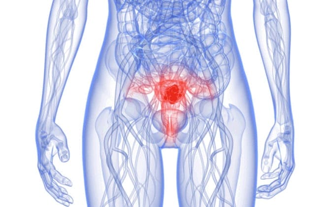
- Poorly differentiated uterine tumors. Development begins in the cervical area, the neoplasm is formed from cells that produce cervical mucus. Glandular epithelial cells form atypical clusters. In later stages, atypical cells quickly spread to the endometrium and muscle tissue of the uterus. The disease is diagnosed during menopause. It is difficult to identify such a tumor, and the same applies to treatment.
- Poorly differentiated prostate cancer. Formed from glandular tissues. The first symptoms appear only at stage 3. In the initial stages, the disease is confused with prostatitis. This type of cancer is difficult to differentiate from squamous cell carcinoma, which has a slower development rate.
- Malignant tumors of the rectum. They are formed from the cells of the epithelial lining of this section of the gastrointestinal tract. They are difficult to diagnose, which is why they are often detected at the terminal stage.
- Lung adenocarcinoma. Develops from the epithelial tissues of the bronchi or alveoli. A lung tumor can be a source of secondary damage to malignant lesions of other organs. This type of cancer develops against the background of genetic disorders that alter the degree of cell differentiation.
- Ovarian tumors. They belong to epithelial types of cancer. The neoplasm consists of highly modified cells and does not have clear boundaries.
- Poorly differentiated colon carcinoma. Improperly developed epithelial cells that make up the tumor produce large amounts of mucus, which accumulates in the intestines in the form of clots.
- Endometrial neoplasms. They develop against the background of benign hyperplasia or excess estrogen. The neoplasm consists of tubular glands lined with ratified epithelium.
Proper nutrition for stomach cancer

This illness is a serious test, accompanied by a complete loss of strength, nausea and vomiting, and intense pain. Treatment is aimed at stopping the growth of the tumor, inhibiting the appearance of new lesions and alleviating the patient's condition. Proper nutrition should also contribute to this.
The general principles can be formulated as follows:
- Reducing the amount of food consumed per day to a minimum. In this case, you are allowed to eat up to 8-10 times a day, maintaining equal intervals between meals.
- Food should be comfortable in temperature - not too hot and not cold, approximately equal to body temperature. This way the body absorbs it better.
- Solid foods should be chewed thoroughly to reduce stress on the stomach.
- Prepare food immediately before consumption.
- Steamed, baked and boiled dishes are acceptable. Fried foods contain carcinogens that irritate the gastric mucosa.
- Nutrition for stomach cancer should promote good bowel movements. This is achieved by increasing the diet of plant foods containing fiber, which improves peristalsis and facilitates the process of releasing the organ. Plant foods contain many vitamins and enzymes that slow down the spread of atypical cells throughout the body.
- Avoid the use of substances that irritate the stomach - hot spices and seasonings.
- Limiting salt intake - the safe amount per day is 5g.
- Reducing fat intake.
A diet for stomach cancer prevents sudden weight loss, makes it easier to tolerate chemotherapy and its side effects, supports the immune system and normalizes metabolism.
One of the reasons for the development of tumors is the fact that modern food products often contain artificial additives (flavor enhancers, flavors and colorings). Such products should be excluded from the menu for stomach cancer forever. A special diet is prescribed about a month before surgery. This is a fractional and regular meal, preferably in the form of mushy, liquid and steam food. In this form, it is better digested and absorbed.