The pancreas is an organ of the abdominal cavity that is responsible for several important functions of the body. The main function is to produce pancreatic juice, which helps break down food, thereby ensuring its better absorption. The formation of stones in the pancreas is a rare disease, but in recent years the incidence of such lesions has become particularly high, especially in Western countries.
A little about the terms
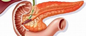
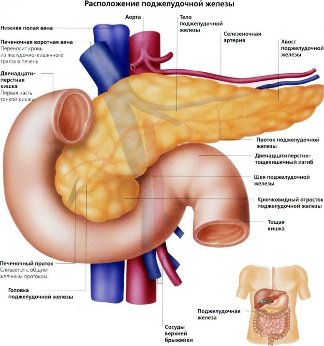
The pancreas or large gland, the most important part of the endocrine system, is located in the upper part of the abdominal cavity and is pear-shaped. Among its functions, a significant place is occupied by the secretion of enzymes, the entry of which into the digestive system ensures the breakdown of products and the absorption of useful and necessary components for the body. Gallstones are the result of crystallization of cholesterol and bilirubin deposits in the presence of pathology in the ducts. Often the provoking factor for their occurrence is pancreatitis; up to 60% of patients with this disease carry stones in the pancreas.
More details about the problem
Stones in the pancreas can have different sizes and shapes. In most cases, such formations are oval, but irregularly shaped calcifications are also common. Most often, such stones form in accessory ducts or in their branches. On the other hand, the formation of solid structures can also be observed in the parenchyma of the pancreas itself.
As for the chemical composition, the main stones of such stones are phosphate or calcium carbonate salts. In addition, such formations may contain compounds of aluminum, magnesium, as well as some substances of organic origin.
Etiology of the disease
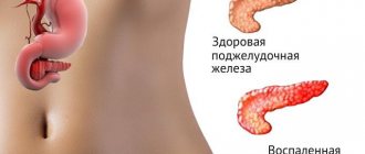
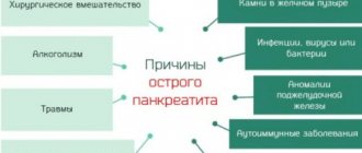
With pancreatitis or other provoking diseases, the normally smooth inner surface of the excretory duct becomes deformed. This leads to stagnation of some of the enzymes, forming a precipitate with its subsequent crystallization into stone. As a result, digestive juices do not completely pass through the duct and become an additional factor that destroys the tissue structure of the organ. The risk of such a complication increases significantly if:
- presence of congenital predisposition;
- sedentary lifestyle and excess weight;
- following a diet with a sharp decrease in body weight;
- liver pathologies and diabetes mellitus;
- blood diseases;
- increased levels of bilirubin, cholesterol in bile juice;
- reaching old age in men;
- gestation;
- taking medications, for example, contraceptives or anti-cholesterol drugs.
To avoid illness, just listen to your body, and if you experience any characteristic discomfort, immediately consult a doctor.
Characteristic symptoms
As a rule, stones in the pancreas are accompanied by symptoms expressed in pain. They can have different durations from short-term ones of a few minutes to long-lasting ones that last for many hours. They differ in varying degrees of intensity, for example, they intensify after eating or when touching the stomach. Places of localization can be:
- upper abdomen or right side;
- right shoulder;
- area between the shoulder blades;
- abdominal area with impact in the back.
In addition to the painfulness of the pathology, the patient may be bothered by frequent nausea, vomiting, bloating, excessive sweating, and uncharacteristic light brown stools.
To treat gastrointestinal diseases, people successfully use Galina Savinova’s method. Read more >>>
Possible complications
The main types of complications remain pancreatitis and diabetes mellitus, or the latter versus the former. However, there is an inverse relationship; inflammation during pancreatitis is also among the pathologies that provoke stones when the ducts in the pancreas become scarred. To maintain hormonal balance, the latter produces insulin, which must leave the ducts without delay to reduce the amount of sugar in the blood. If this does not happen, insulin deficiency and ultimately diabetes mellitus occurs. With prolonged blockage, acute pancreatitis is formed, which is accompanied by:
- high temperature;
- long-term pain;
- infections.
In addition to the acute pain caused by the blockage, yellowness of the skin and a characteristic change in the color of the eyeballs are added. Severe symptoms will most likely require hospital treatment, and there is a high probability that surgery will be necessary.
About symptoms and possible complications
A typical symptom of the disease is prolonged, severe pain in the upper abdomen or right side. Painful sensations can last from fifteen minutes to several hours. They can also be felt between the shoulder blades and in the right shoulder. Sometimes patients complain of nausea and excessive sweating.
Attacks of pain can be separated by weeks, months or even years. Stones can also lead to attacks of acute pancreatitis. Signs and symptoms vary depending on the person. These may include:
- pain in the epigastric region;
- pain in the abdomen that radiates to the back;
- abdominal pain that gets worse after eating;
- nausea;
- vomit;
- light brown stool color;
- sweating;
- increase in abdominal size;
- painful sensations from touching the stomach.
In addition to blocking the release of digestive enzymes, stones can cause serious health problems: the pancreas produces hormones necessary to control sugar levels in the body. If these hormones are not released or are released insufficiently, diabetes occurs.
Attacks of severe pain can occur every day for a long period
If the blockage lasts longer than a few hours, inflammation can occur—a condition called “acute pancreatitis,” which can cause high fever, prolonged pain, and ultimately lead to infection of the gland.
The pain is a consequence of the blockage of the gland duct.
Bile duct stones usually cause pain, fever, and bile leakage (yellowing of the eyes and skin), sometimes accompanied by itching. If your symptoms are severe, you may need to be hospitalized.
Most often it is required for surgery, patient monitoring and treatment with antibiotics and painkillers.
Diagnostic options
Stones in the pancreas can be easily detected by radiography, which is used for an overview of organs in the abdominal cavity. Concretions are highlighted with multiple or single rounded shadows. Visually localized below the urinary process, on the right and left sides relative to the midline. However, the results of this method are usually not enough; to clarify them, you will have to do:
- Ultrasonography, which will allow you to evaluate the location, size, outline and structure of formations.
- Magnetic resonance imaging, with the help of which a three-dimensional model of the organ and its vessels is reconstructed with a detailed display of defects.
- Retrograde cholangiopancreatography, clarifies the localization of pathological formations. It also determines the patency of the duct.
Based on the results of diagnostic procedures, a gastroenterologist is consulted, who determines an individual treatment regimen for the patient.
Reasons for formation
The process of stone formation is quite long. The trigger can be:
- Inflammatory disease in the gastrointestinal tract, especially chronic.
- Failures in metabolic processes.
- The presence of stagnation.
- Chronic alcoholism.
- Bad habits.
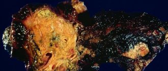
Photo of stones in the pancreas
Under the influence of irritating factors, the gland juice becomes thicker. This leads to the fact that calcium salts actively begin to be deposited. The cause may also be a disease that occurs without symptoms. These can be cysts, small benign neoplasms.
Types of therapy
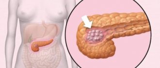
Even in an asymptomatic course of the disease, stones pose a danger to the body. When neglected, they provoke various pathologies of the pancreas, have a negative impact on the general condition, and can cause oncology of the epithelium. For this reason, it is necessary to be treated in all cases. For small lesions, conservative techniques are usually used.
However, their limited effectiveness makes it necessary to use the removal of large pancreatic stones in accordance with individual indications. These include:
- endoscopic technique;
- pancreatomy;
- external shock wave lithotripsy;
- laser version of lithotripsy.
Any operation listed above will allow you to effectively remove stones in the pancreas; however, an important condition for success is the qualifications of the surgeon and strict adherence to the procedure. Consequences may include postoperative pain symptoms, hematomas, and incomplete removal.
Methods of conservative treatment
They are used when pathology is detected in the primary stage. The use of medications is combined with dietary nutrition and sanitary spa treatment. Taking prescribed medications should neutralize inflammatory processes, reduce swelling in the affected area, and normalize metabolism. Enzyme deficiency is compensated by taking enzyme-containing drugs. The result may be migration of stones into the intestines with subsequent removal to the outside, or their dissolution when taking tablets containing chenodiol and ursodiol.
Endoscopic removal
A tube with a camera is used to perform the operation. To detect the formation, it is introduced into the stomach through the pharynx, after which the stone is removed. The operation is almost painless and does not require a long recovery period. But for large or group formations it is useless.
External shock wave lithotripsy
A crushing method that uses directed sound waves. The growths are crushed to the state of sand. Then the remains are removed independently or removed endoscopically. The operation requires general anesthesia. Rehabilitation does not take much time.
Laser method
An atraumatic technique, which is carried out without violating the integrity of tissues, is the use of a laser. A micro-camera is used for detection, and the video image is displayed on the monitor. The manipulator is inserted into the body, after which the calculus is broken into sand and comes out naturally. The procedure is delicate, leaves no scars and minimizes the risk of relapse. It has almost no complications and is highly effective.
Minimal trauma during the removal process ensures rapid recovery of the body.
Surgery
For multiple large lesions, pancreatotomy is used. The growth is removed through an incision. If the pathology is multiple, it is performed on the entire surface of the organ. The inside is completely cleaned, including the smallest particles. The operation is technically complex, often causes complications, and is performed exclusively in extreme cases when other methods are unavailable or contraindicated.
Application of total pancreatomy
Used for oncology, it involves the removal of the gland and part of the duodenum. Indications also include relapse of chronic pancreatitis and total pancreatic necrosis. The consequence is the occurrence of exocrine and endocrine insufficiency. Digestive function is impaired and diabetes mellitus develops. Lifelong use of enzyme-containing drugs and insulin is necessary, which compensates for the lack of natural secretion.
Treatment methods
If there are stones in the pancreas, the following can be used:
- surgery,
- conservative therapy,
- folk remedies,
- diet.
Operation
This method is used for the following indications:
- Frequent and prolonged pain attacks.
- Long term illness.
- Progressive inflammatory process.
- Exhaustion of the body.
One of the treatment methods is ERCP. Small stones are removed from the organ using an endoscope. If the formation is large, then a small incision is made in the duct, followed by pushing the formation into the intestinal section.
The ESWL method is considered atraumatic. With it, stones are crushed to a powder using remote shock wave lithotripsy. After this, the powder is removed from the body independently or with the help of an endoscope.
The procedure is done under general anesthesia. The patient is placed on his stomach, which touches the emitter. Sometimes the operation is associated with pain. Bruising may occur.
Sometimes diffuse tissue calcification is detected during surgery. Then a total pancreatectomy is performed. It involves complete removal of the organ. Since exogenous and endocrine insufficiency develops after such manipulation, the patient must receive treatment throughout his life.
Conservative therapy
This method of exposure is selected depending on the size and number of stones and the patient’s condition. In some cases, it is possible to use special preparations that dissolve such formations.
Such products can only cope with small stones. These include Henodiol, Ursodiol, which make bile more liquid, promoting the destruction of stones.
Medicines that affect metabolism are also prescribed. If its violation led to the formation of stones. Throughout the course you must follow a daily routine.
Folk remedies
Treatment with this method cannot occur without the consent of a doctor. Herbal medicine helps with stones:
- Pour crushed chicory root into water and boil for 5 minutes. After cooling, the drink is filtered and drunk in small portions throughout the day.
- A tablespoon of barberry bark is poured into 250 ml of boiling water. It is necessary to cool the broth for 20 minutes. Take a tablespoon several times a day.
- Burdock root is ground to a powder. You need to take 2 tablespoons. This amount is brewed in 0.5 liters of boiling water and left for 3 days. You need to drink a filtered drink 30 minutes before meals throughout the day.
To stimulate the functioning of the organ, you can drink tea from linden, chamomile, violet, mint and St. John's wort. All components are taken in equal quantities. A glass of drink requires 50 grams. mixtures. You need to drink 2 times a day.
Diet
Following a diet is an excellent prevention of attacks. It makes it possible to reduce the severity of symptoms and slow down the growth of stones. The diet involves reducing the calorie intake. If you break this rule, then there is a high probability of stones moving along the duct.
This may cause an attack.
- When doing diet therapy, several rules must be followed:
- Eliminate fatty, sweet, spicy foods.
- Eat in small portions so as not to put stress on the pancreas.
- Lead an active lifestyle, engage in physical therapy to improve motor skills.
- Keep your body in shape by using dietary supplements.
Try to reduce your consumption of eggs and oils. The diet should include stewed or boiled dishes from vegetables, meat and fish. A special diet allows you to eat pasta and dairy products.
Preventive measures
There are no radical measures that can prevent the development of pathology, especially in the presence of favorable factors. The risk of its occurrence can be reduced by:
- dietary nutrition;
- rejection of bad habits;
- self-monitoring of the body’s condition and the development of symptoms.
The earlier inflammation is detected and appropriate measures are taken, the greater the chances of avoiding the occurrence of growths and the need for their surgical treatment. Even if surgery cannot be avoided, performing it in the early stages will prevent the use of radical measures.
The pancreas is one of the important organs responsible for the production of gastric juice so that food is better broken down and absorbed. Often the main duct of the gland has a smooth and even structure; through the duct, the juice is directed into the small intestine. When pancreatitis occurs, a modification of the duct occurs; in some areas it becomes narrow due to the presence of an inflammatory phenomenon in it. Therefore, the juice does not come out in full and stones form in the pancreas. When the duct is blocked, the patient experiences unbearable pain that requires immediate treatment.
Reasons for education
The presence of stones in the pancreas is a rather rare disease, but recently the number of victims has increased significantly.
The procedure for forming pancreatic stones is complex. To start it, a combination of a number of exciting reasons will be required. The procedure for the development of stones is organized on the retention of digestive juice, which thickens as it accumulates. Then the formation of a protein mass occurs with the gradual development of the phenomenon of its calcification. Formed stones in the pancreas contribute to pressure on the ducts, leading to necrotic changes.
The reasons that contribute to the formation of stones are not fully understood. However, there is a combination of reasons that can increase the risk of developing the disease.
- Inflammatory diseases in the stomach and intestines - pancreatitis, cholecystitis, duodenitis.
- Narrowing of the gland ducts due to the presence of a tumor formation, whales.
- Age – patients over 40 are susceptible to the formation of stones in the organ.
- A metabolic disorder is a violation of the absorption of calcium and phosphorus.
- Infectious diseases.
- Excess weight.
- Improper lifestyle – drinking alcohol, smoking, poor nutrition.
- Hormonal imbalances.
At risk are patients who have various painful phenomena in the abdominal cavity, especially when they obstruct the outflow of digestive juice.
Stages of stone formation:
- At the stage of development of the disease, stones appear. The reason is the thickening of digestive juice. The victim has an insoluble mass of protein localized in the ducts.
- The second stage of the disease is characterized by the separation of calcium salts, which leads to a worsening of the situation.
- At the third stage, an infection occurs, after which stones form in the gland.
Stages of stone formation in the gland
Stones do not appear instantly, but gradually.
At the first stage, pancreatic juice is concentrated, which is then converted into an insoluble protein mass.
The second stage consists of the accumulation of calcium and phosphate salts in the damaged secretion, due to which the chemical composition changes.
The third stage is caused by the appearance of an infectious disease, as a result of which all the signs of stone formation appear
Symptoms of pancreatic stones
The severity of symptoms of stones in the pancreas depends on the stage of the stone. The formations are located in the ducts or parenchymal part of the organ.
In all situations, if pancreatic stones have formed, the symptoms initially manifest as unbearable pain. Painful discomfort lasts from 2-3 minutes to several hours, the pain moves to the right shoulder and the area in the middle of the shoulder blades. The pain is burning, sometimes manifesting as colic, affecting the abdominal area and lower back. Attacks occur every day or in rare cases (once a month, a year).
Stones in the pancreas are characterized by the following manifestations:
- severe pain during meals when you touch your stomach;
- nausea;
- vomiting bile;
- stool takes on a light color and contains undigested fat—fatty bowel movements;
- There is a lot of saliva;
- outwardly the stomach appears swollen;
- weakness;
- sweating
In addition, if there are stones in the pancreas, the work of enzymes is blocked, and therefore the victim has serious problems.
The pancreas is responsible for producing hormones that control glucose levels in the circulatory system. Due to existing stones, the excretion of hormones may decrease, which will cause the development of diabetes. In this case, the victim is recommended to undergo testing for this disease.
If, due to stones, long-term intussusception of the ducts develops, sometimes inflammation of the pancreas begins, which is characterized by an acute course of pancreatitis. This phenomenon leads to an increase in temperature, prolonged pain and organ poisoning. Often, painful discomfort develops due to the inability of fluid to pass through the ducts.
Stones forming in the bile duct lead to excruciating pain, fever and skin jaundice, indicating that bile has formed. If such signs are observed, the patient should urgently seek medical help. In one situation out of 20, pancreatic stone disease occurs without obvious signs. This phenomenon causes difficulties when diagnosing pathology.
Stages of the disease and their symptoms
The process of formation of stones in the pancreas occurs in several stages. Each stage has its own symptoms and causes of pain in the pancreas.
At the initial stage, under the influence of factors such as smoking, alcohol, metabolic and hormonal imbalances, pancreatic juice thickens, and protein fractions fall out of it. They form protein plugs into which calcium salts penetrate. The formation of sand grains in these deposits begins.

The characteristic symptoms of sand in the pancreas are acute and persistent pain in the stomach. The pain radiates to the shoulder blades and right side of the back, nausea and vomiting occur. Pain occurs after eating or drinking alcohol.
If obstruction of the pancreatic ducts persists for a long time, calcified stones grow to the size of stones. They block the gland duct and cause inflammation, which is often accompanied by infection. Pain occurs because pancreatic juice cannot leave the pancreas.
Symptoms of stones in the pancreas:
- heat;
- yellowed skin and whites of the eyes;
- pain lasts from several hours to several days;
- the patient develops steatorrhea (oily, foul-smelling stool);
- a person loses weight.
The patient requires urgent hospitalization to monitor his condition, treat him with antibiotics and painkillers, and sometimes for surgery.
Diagnosis of pancreatic stones
It is difficult to make a correct diagnosis based only on external manifestations, because similar symptoms of an inflammatory disease also indicate other pathologies.
As a result, in order to clarify the presence of stones in the pancreatic ducts, the following measures are carried out:
- laboratory tests, including biochemical blood tests, enzyme studies;
- Ultrasound – allows you to examine the echogenicity of the organ, its size, and identify the presence of stagnant processes in the ducts;
- MRI – helps to accurately determine whether stones are present or absent in the pancreas;
- endoscopic examination - they take biomaterial and study the density of the organ;
- retrograde cholangiopancreatography – clarifies the placement of stones, assesses the stage of duct patency.
Often in gastroenterology, diagnosing stones is not difficult. In gastroenterology, during radiography of the peritoneum, stones in the organ are visible as small round shadows, in a single or multiple copy. Stones are localized under the sternum, on the left and right sides of the center. To clarify the diagnosis, photographs are taken in various projections.
Diagnostics
It is difficult to make a correct diagnosis based only on external signs, since such symptoms may accompany other pathologies. Therefore, for clarification, it is prescribed:
- Ultrasound. Shows the echogenicity of the organ and its size. The method makes it possible to determine the presence of stagnant processes in the ducts.
- MRI. Makes it possible to accurately determine the presence or absence of stones.
- X-ray. Displays the size of formations, their location and ability to move.
An effective method is endoscopic examination, when an endoscope is inserted through a small hole to examine the organ and take biomaterial to study the density.
Additionally, laboratory tests are performed. If increased activity of produced enzymes is diagnosed, this indicates that there is acute inflammation. To identify them, blood, urine or feces are used.
Treatment of pancreatic stones
Victims with stones in the pancreas are treated by a gastroenterologist. The procedure for pathological measures will depend on the volume of formations, their structure and size. Methods for treating stones in the pancreas.
- Conservative measures.
- Operational method.
- Use of folk remedies.
- Diet food.
Medication route
Despite the complexity of the condition, treatment procedures begin with conservative methods.
- Relieve inflammation.
- Stop swelling of the gland and ducts.
- Establish metabolic processes.
Patients are prescribed enzymes - Pancreatin, the daily dosage of the drug is selected by the doctor. Sometimes it is possible to use special agents that can dissolve stones in the pancreas. Such drugs are able to overcome small formations, making bile much lower and destroying the formations.
Against the background of conservative treatment, the victim’s well-being can significantly improve; formations of insignificant size can themselves move into the intestinal area.
Surgical solution to the problem
If large formations are present, it is often impossible to cure using conservative methods. In this situation, surgical intervention is required. Removal of stones in the pancreas is carried out if:
- frequent and prolonged pain occurs;
- in the presence of a chronic inflammatory phenomenon;
- developing inflammation;
- exhaustion of the body.
One of the means of surgical intervention is ERCP. Small formations are removed using an endoscope. If there are large stones, an incision is made in the duct and the stone is pushed into the intestinal area. Endoscopic removal is better tolerated by patients; such a cure does not require a long adjustment, and it is impossible to remove all formations in this way.
What treatment plan can the doctor prescribe?

Patients who have stones in the pancreas should visit a gastroenterologist.
The treatment itself consists of initially eliminating the inflammation process, normalizing the metabolism of calcium and phosphorus, and also reducing swelling of the pancreatic tissue. The doctor must first prescribe enzyme treatment and prescribe a strict diet.
By adhering to all the doctor’s recommendations and following a diet and rest regimen, the patient may feel improvements, and small stones found may move into the intestines on their own.
In addition, you can use special medications (ursodiol, chenodiol) that will help dissolve stones in the pancreas. Their action is aimed at liquefying bile and destroying the stone itself. However, this method will be effective only for small stones, which mainly contain cholesterol.
If large stones are found, then traditional methods will not help. In this case, surgical intervention will be required, which is characterized by damage to the integrity of the affected organ and removal of the cause of the disease. Surgical intervention is used when attacks become frequent and the body cannot cope.
Is it possible to remove stones from the pancreas without surgery? Nowadays, medicine is opening up new possibilities and methods for treating various diseases every day.
Removing stones from the pancreas with a laser is one of the methods of modern medicine. The procedure is as follows: sound waves are used, under the influence of which it is possible to crush or crush stones into powder. And the remaining particles can come out on their own, or they can be eliminated using ERCP.
Prognosis and prevention
The prognosis and prevention of stones depend on the severity of the pathology and the presence of accompanying diseases. Often, if the pancreatic stone is removed in time and treatment begins, the prognosis is favorable.
In 85% of surgical treatment situations, it results in an improvement in well-being. Only 2% of patients die after surgery.
There are no specific preventive measures for the formation of pathology. It is possible to reduce the risk of stone formation by following a gentle diet, not smoking and not drinking alcohol. Patients who are genetically predisposed to diseases of the stomach and intestines should promptly consult a doctor if they have various symptoms regarding the digestive system.
When a stone is discovered in the pancreas, it is dangerous and requires a quick response. If the pathology is not treated, there is a threat of malignant formations in the pancreas gland. Surgical treatment will get rid of the disease in a short time. After removing the stones, you need to follow the doctor’s recommendations and a healthy lifestyle, then the threat of relapse will be minimal.
The pancreas is an organ of the abdominal cavity that is responsible for several important functions of the body. The main function is to produce pancreatic juice, which helps break down food, thereby ensuring its better absorption. The formation of stones in the pancreas is a rare disease, but in recent years the incidence of such lesions has become particularly high, especially in Western countries.
Diagnosis and treatment
Sugar level
Man
Woman
Enter your sugar or select your gender to get recommendations
Pancreolithiasis refers to diseases of the digestive system, ICD code - K00-K93. An X-ray of the abdominal organs is performed. It allows you to visualize stones, which appear as a small round shadow. There are single and multiple. More accurate diagnostic methods include CT and MRI.
The patient definitely needs to consult a gastroenterologist. Treatment always begins with medication. It is aimed at stopping inflammatory processes, designed to normalize the exchange of calcium and phosphorus in the body, reduce swelling of the pancreas and main duct. Anti-inflammatory and painkillers are prescribed.
Enzyme therapy is prescribed, special attention is paid to nutrition, and diet number five is prescribed. If the stones are small, then tablets can help improve the patient’s condition; small stones will pass into the intestines on their own.
If you have large stones, there is no point in taking medications to dissolve them. Surgery is required because there is a danger to health and life. In medicine, minimally invasive methods have been significantly modernized. For example, endoscopic removal of main duct stones is often used. A puncture is made and stones are removed using an endoscope.
Advantages of the endoscopic method:
- Lack of long-term rehabilitation.
- Minimum complications after the intervention.
The disadvantage is that the manipulation may not remove all tumors in the pancreas. If there are a lot of large stones that are localized in the body and tail, then it is better to use the laparotomy method.
Surgical treatment can be carried out using the crushing method. The operation involves a shock wave effect on the stones, which allows them to be crushed to the smallest grains of sand. They subsequently leave the body during bowel movements.
A modern method is laser stone removal. The stones are visualized using a video camera, and the image is transmitted to the monitor. Next, targeted breaking is carried out to the state of sand. Reviews from doctors note that laser treatment has many advantages. They have a high degree of fragmentation, minimal trauma, a short recovery period, and no scars.
What is the cost of laser removal? The price is determined by the pricing policy of the medical institution, the qualifications of the doctor and other aspects. On average, the starting cost is from 15,000 rubles.
The effectiveness of any intervention depends on the qualifications of the doctor. Complications include pain, hematoma in the surgical area, and incomplete removal of stones.
Features of the disease
The main duct of the pancreas, through which the juice goes to the small intestine, most often does not have any irregularities and is smooth. In people who suffer from chronic pancreatitis, the main duct in the pancreas has an irregular shape: in some places it is very narrowed. This happens due to scarring and regular inflammation in the gland. As a result of this, the juice is not completely eliminated; in 22-60 percent of patients with this disease, stones begin to actively form in the stomach. When the duct is blocked, they cause acute, severe pain.
Most often, the formation of stones occurs from chronic inflammation and changes in metabolism. They are created due to the deposition of calcium salts in the gland and can block the production of enzymes that perform digestive functions.
In addition to stones, stones that occur in the gall bladder form in the pancreas. They can accumulate in the bile duct, which is combined with the pancreatic duct.
A gallstone is part of a solid substance. It forms when components of bile, including bilirubin and cholesterol, precipitate and create crystals.
If any of these stones block the common duct, then the digestive enzyme begins to act on the gland itself, thereby destroying its tissue.
Stones in the pancreatic duct can be as small as sand, or they can be much larger. It has not yet been established why some people experience them and others do not. But there are a number of reasons that increase the chance of their formation :
- excessive excess weight;
- diabetes;
- liver pain;
- a large amount of cholesterol or bilirubin in bile;
- age over forty years;
- lack of sports and necessary mobility in life;
- Women are most prone to this phenomenon;
- a tendency to form gallstones at the genetic level.
Cholesterol stones and bilirubin components occur most often in the following categories of people:
- patients who suffer from severe liver diseases;
- women over the age of twenty, this is especially common in pregnant women;
- patients who use certain medications, including birth control pills and cholesterol-lowering pills;
- men and women who are susceptible to obesity;
- patients with blood diseases. For example, leukemia or sickle cell anemia;
- men over the age of sixty;
- people on a demanding diet, in which weight begins to be lost in a short period of time.
General information about pathology
The appearance of stones in the pancreas is a rather rare pathology that occurs as a result of the development of a chronic form of pancreatic disease and sudden changes in metabolic processes. Intense deposition of calcium salts in the pancreatic cavity promotes the formation of stones, which block the release of enzymatic components produced by the gland, which leads to their activation in the pancreatic cavity and the development of a necrotic process against the background of the self-digestion of this organ.
Therefore, timely detection of stone formation in a parenchymal organ can not only give a positive result in treatment, but also improve digestion processes, improve the patient’s quality of life and prevent the likelihood of death.
In addition to those stones that form in the pancreatic cavity, there are also those that can form in the gallbladder cavity, causing the development of cholelithiasis. The formation of stones in the lumen of the gallbladder or in its ducts contributes to the development of an inflammatory process of an acute or chronic nature, called calculous cholecystitis.
The development of the acute form of calculous cholecystitis occurs due to the prolonged course of gallstone pathology, which, against the background of prolonged obstruction of the bile duct, causes the development of inflammation of the walls of the gallbladder. This is due to the fact that infectious microorganisms from other organs penetrate into its cavity against the background of a decrease in the antiseptic properties of bile. At the same time, the development of infection can cause disruption of the muscular membranes of the bladder itself or its ducts, causing dysfunction of the biliary tract.
The formation of stones in the pancreatic cavity does not occur in every patient with the development of cholelithiasis, or chronic pancreatitis. Their formation mechanism has not yet been thoroughly disclosed, but many experts identify a number of provoking factors, in the presence of which the likelihood of stone formation increases several times:
- increased concentration in bile of a substance such as bilirubin,
- a large number of extra pounds,
- sedentary lifestyle,
- age indicator, usually stones begin to form in people over 40-45 years old,
- development of diabetes mellitus,
- pathological changes in liver functionality,
- hereditary predisposition.

The formation of bilirubin, or as they are also called cholesterol, stones in the gall bladder occurs most often in the following categories of people:
- in people with severe liver or blood disease,
- in girls over 20-25 years old, especially during pregnancy,
- in the strong half of humanity who are over 60-65 years old,
- if you have a lot of excess weight,
- for those who go on a strict diet regimen and lose a huge number of kilograms in a short period of time,
- from taking contraceptives and cholesterol-lowering medications.
Let's take a closer look at how cholelithiasis and inflammation of the pancreas are connected.
As already mentioned, the pancreas and gallbladder are interconnected by being close to each other and performing similar functions. Indeed, the pancreatic duct of the gland flows into the same opening of the duodenum as the duct of the gallbladder in the area where the nipple of Vater is located. In most cases, both ducts merge into one and flow into one opening of the nipple, but there are cases when each duct flows into a separate opening of the duodenum, then the likelihood of interaction between the two ducts is minimized.
When two ducts merge, which happens in 75% of cases, a complication arises with cholelithiasis in the form of exacerbation of pancreatitis.
The merged two ducts interact with each other on a constant basis. Therefore, when a stone is pushed out of the gallbladder cavity, it passes freely through the bile duct and gets stuck at the confluence of the ducts in the area of the Vater nipple, since this area is the narrowest point of passage into the intestinal cavity among all bile ducts.
The production of pancreatic enzymes by the pancreas and bile by the liver remains at the same level. Therefore, these fluids, passing through the ducts, do not have the opportunity to exit into the intestines to take part in the digestive processes, since the passage to the intestines is blocked by a stone. This leads to increased pressure in the ducts, which causes them to rupture. All the fluid contained in the ducts enters the pancreatic tissue.
Pancreatic juice, consisting of enzymes that break down proteins, fats and carbohydrates, while in the cavity of the duct, is inactive. But when the walls of the duct rupture, it is activated, which causes intensive development of diffuse changes in the pancreas, the beginning of processes of self-digestion, the appearance of an attack of acute pancreatitis, or the development of such a serious and life-threatening pathology as pancreatic necrosis.
Reasons for development
The basis for this process of stone formation is a violation of calcium metabolism in the body. Normal calcium metabolism will depend on several factors:
- disturbances in the hormonal system;
- nutritional features;
- disturbances in the process of calcium absorption.
Local infectious and inflammatory processes that occur in the vicinity of the pancreas can affect the damage caused by pancreolithiasis. Due to inflammatory processes in the ducts and disruption of the epithelium, detachment of some cells occurs, which negatively affects the functioning of the body and disrupts the overall biochemical composition of the pancreatic secretion.
The excretion of pancreatic juice slows down several times in the presence of:
- pancreatitis;
- duodenitis;
- cholecystitis;
- difficulties in the functioning of the sphincter of Oddi;
- stones in the lumen of the bile duct;
- post-infectious edema of the sap.
The processes of tumor and cyst development also in most cases provoke the appearance of pancreolithiasis. Very rarely, the process of deposition of kidney stones will occur after syphilis or appear after constant smoking or drinking alcohol. The components that are found in alcoholic drinks and cigarettes negatively affect the composition of the secret fluid of the pancreas and provoke an increase in their calcium content.
Increased levels of calcium in the body result in pancreatic juice thickening and causing stone formation. This process has three stages :
- At the first stage, the amount of juice increases significantly. It becomes somewhat denser. Once it enters the ducts, it changes to an insoluble protein mass.
- At the second stage, a strong thickening of the ring flakes occurs, which include salts in their composition. This continues to change the overall composition of the secretion towards the precipitation of sedimentary inclusions.
- In the end, a persistent infection appears and obvious symptoms of the disease develop.
Stones can get stuck in the lumens of the duct, and also form in the parenchyma of the gland. Immediately after their appearance, their overall structure has a less dense shape, but over time it begins to become harder.
pancreatic stones
- excessive weight;
- in old people;
- when leading a sedentary lifestyle;
- liver diseases;
- affected by diabetes mellitus.
This disease most often affects women, especially the age group after forty years.
Causes and symptoms
A person is at risk of developing stones in the pancreas when he is diagnosed with:
- inflammatory diseases of the digestive tract;
- cholelithiasis;
- hormonal imbalances;
- pathologies of the abdominal organs that impede the outflow of pancreatic juice;
- disturbance of calcium-phosphorus metabolism;
- changes in the chemical composition of pancreatic secretions against the background of infectious diseases;
- bad habits.
At the early stage of stone formation, pancreatic juice thickens. When it enters the ducts, it turns into an insoluble protein mass. Next, calcium salts are deposited, which thickens it even more and changes the chemical composition. The next stage is characterized by the addition of infection and the appearance of symptoms of pancreolithiasis.
The manifestation of pancreolithiasis depends on the location of the stones, accompanying diseases, and the degree of inflammation. In accordance with this, there are 2 variants of the disease:
- stones are localized in the ducts;
- calcium salts are located in the parenchyma.
This division is conditional, since in most cases both variants of the disease are combined. Diagnosis of pancreolithiasis is difficult, since there are no specific symptoms directly indicating the presence of stones. The main symptom of the disease is pain. With pancreolithiasis, they are most often localized in the epigastric region and can radiate to the lower back and between the shoulder blades.
The intensity of pain varies and is paroxysmal in nature. The pain may subside between attacks, intensify with a new attack, and be accompanied by nausea and vomiting. Discomfort may occur due to stress or poor nutrition. Due to disruption of the pancreas, there is a risk of developing diabetes. Symptoms indicating the presence of stones in the pancreas:
- yellowing of the skin;
- increased secretion of saliva;
- nausea, vomiting with bile;
- fat drops in the stool;
- the presence of stones in the stool.
Sometimes a stone or sand from the gallbladder moves along the bile duct to the main duct of the pancreas, provoking the appearance of an inflammatory process in it. Then they talk about the development of gallstone pancreatitis.
Stones in the pancreas: symptoms
At the initial stage of development, a person may not even be aware of the onset of the disease, since at this time the formation of calcifications does not at all affect the patient’s well-being and does not impede the functioning of the digestive system. After their significant increase, pressure on such an organ increases, which as a result leads to the appearance of the first discomfort, which is more like the symptoms of pancreatitis.
Very often, the first signs of the disease occur after severe overeating, after consuming fatty foods or alcohol. In this case, in the abdomen one can note girdling spasmodic pain sensations of a burning nature, which are more like colic. Moreover, such manifestations occur along with a feeling of nausea and vomiting with a hint of bile, and sweat may appear on the body. Pain may extend to the right side, shoulder, shoulder blade or collarbone. During and after an attack, undigested or undigested fat can be found in the patient’s stool, which looks more like white inclusions.
The first attacks of the disease can be widely separated in time; in some cases, not only months, but even whole years can pass between them.
With the development of greater blockage of the gland duct by calcifications, exacerbation attacks occur regularly. The symptoms may differ slightly, depending on the specific location of the stones in the gland structure. In some cases, the disease does not manifest itself in any way until complications develop. In other cases symptoms as:
- heartburn;
- attacks of fever;
- strong secretion of saliva;
- vomiting with bile;
- the skin and sclera begin to turn yellow;
- feces become lighter;
- several hours after eating food, pain occurs in the abdominal cavity, which then radiates to the lower back or subscapular region.
Blood tests of such a patient can determine leukocytosis, increased glucose levels, a large number of enzymes in the liver, as well as the pancreas. Based on these indirect symptoms, the disease can be detected by chance during a periodic examination.
As the stage of the disease worsens, necrosis of individual parts of the pancreas occurs, the number of enzymes and overall secretion decrease. Very often, such changes occur together with a disruption in the production of hormones that control blood sugar levels, which can result in diabetes.
If the stones begin to move, they can get stuck in the flow, thereby closing the lumen, resulting in severe blockage and obstructive jaundice, which will lead to severe yellowing of the patient's skin.
If such stagnation occurs over several hours, then the bile has a dangerous effect on the parenchymal tissue of the gland. This can develop inflammation, and in special cases, provoke an abscess, peritonitis or bleeding.
In case of particularly severe symptoms, the patient should not wait for the situation to worsen, but urgently call an ambulance, which will hospitalize the patient.
Treatment with folk remedies
Folk recipes will help relieve severe symptoms in chronic pancreolithiasis and even dissolve stones:
- A tablespoon of crushed barberry bark is steamed with a glass of boiling water and kept at low heat for a quarter of an hour. Filter the decoction and take a tablespoon warm three times a day;
- Burdock root is ground and 2 tablespoons of powder are steamed in half a liter of boiling water. After 3 hours, filter and drink during the day half an hour before meals.
- Steam a teaspoon of chopped blueberry leaves in a glass of boiling water and leave for 45 minutes. You need to drink the infusion throughout the day, taking one sip at a time.
- Mix 20 g of Icelandic moss and a glass of apple cider vinegar. Infuse for 2 days, filter and take a teaspoon of infusion, previously diluted in a glass of warm water, for two weeks.
- A teaspoon of crushed dandelion roots is mixed with 3 teaspoons of mint. The mixture is boiled for 7 minutes in half a liter of water and left for 30 minutes. Take 50 ml before meals.
Carrying out treatment of the patient
Therapy for stones in the pancreas always includes a complex effect. The main measures are aimed at:
- quickly eliminate pain and other symptoms;
- elimination of calcifications;
- establishing metabolism in the body;
- restoration of the amount of enzymes;
- relief of pain and other symptoms;
- preventing the re-development of calcifications.
Methods of combating calcification in the pancreas in this case are divided into surgical treatment and conservative treatment.
Diagnosis of the presence of stones in the pancreas
The examination of the patient begins with palpation; in the acute stage, the following are usually noted: tension of the abdominal muscles, pain when pressed, high sensitivity.
A complete blood count reveals an elevated white blood cell count and an increase in the erythrocyte sedimentation rate (ESR), which indicates an inflammatory process. High activity of the amylase enzyme is found in blood serum and urine; the higher it is, the more dangerous the pathology.
Hardware research methods are the most informative and help to differentiate the presence of stones in the pancreas from other pathologies with similar symptoms. Used: radiography, ultrasound, computed tomography.

Elimination without surgery
This method helps to achieve the desired effect with small stones and with rare attacks of the disease.
To begin with, conservative treatment requires getting rid of the pathology that led to the formation of stones in the stomach, eliminating inflammation, establishing the general exchange of phosphates and salts in the patient’s body, and also reducing swelling of the gland parenchyma and ducts.
For this, the patient is prescribed the following drugs:
- choleretic agents;
- secretory medications;
- antibiotics in case of infectious inflammation;
- antispasmodics, as well as antibiotics to relieve pain.
At the moment when the general condition of the patient improves, attempts begin to move calcifications from the gastric duct and remove them into the intestines. For this purpose, drugs are used that can relieve ductal hypertension.
Surgical intervention
Many patients do not know whether the pancreas is being operated on. Surgery is permitted when the following lesions exist:
- long-term disease with severe and regular attacks;
- serious condition of the patient;
- insufficiency in the pancreas;
- It is impossible to eliminate pain with standard painkillers.
There are several types of surgery. In each case, the type will be selected individually, based on the overall size and location of the stones, the condition of the patient and the equipment in the hospital:
- The most modern and less invasive method is considered to be lithotripsy, which involves crushing calcifications under the influence of shock wave energy. At the end of crushing, the fragments or powder are removed from the gland on their own, but if this does not happen, then they are removed forcibly. The advantage of this technique is that it is performed on an outpatient basis. The procedure lasts thirty minutes and is performed under general anesthesia.
- A minimally invasive procedure is endoscopic retrograde cholangiopancreatography. During this operation, a special endoscopic tube is inserted into the digestive tract, at the end of which there is a camera. With its help, a specialist monitors deposits in the gland, while eliminating them. This method has a shorter recovery time and is much easier to tolerate by the patient, but if there are a large number of deposits and their large size, the endoscopic method will not be effective.
In rare cases, a bypass procedure is performed to create a bypass from the glands to supply pancreatic juice to the duodenum. But only a qualified attending physician can perform this procedure. It must be remembered that the liver is a unique organ on which well-being and health will directly depend, so it is very important to treat it promptly and correctly.