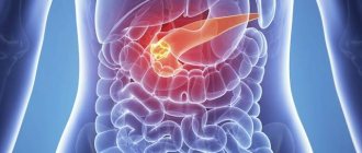
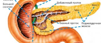
Causes of development of chronic pancreatitis
- Chronic pancreatitis is a persistent inflammation of the pancreas with irreversible morphological changes, usually associated with pain and malabsorption.
- Prevalence of chronic pancreatitis 27:100,000
- Analysis of epidemiological data shows marked geographic variability, which may be due in part to differences in drinking patterns.
Causes:
- The most common cause of chronic pancreatitis is alcohol abuse (70-90% of cases);
- However, only 5-15% of people who regularly drink large amounts of alcohol develop chronic pancreatitis;
- Smoking contributes to the development of calcific pancreatitis;
- Other forms are tropical pancreatitis, hereditary form, metabolic forms, pancreatitis due to obstructive causes, and idiopathic pancreatitis.
When to do it?
For MRI of the pancreas, indications may be as follows:
- Girdle pain in the abdomen, periodic pain in the stomach and hypochondrium, especially some time after eating
- Digestive problems (diarrhea, difficulty digesting fatty foods, sour belching, heartburn, etc.)
- Cancer search and detection of metastases
- Suspicion of pancreatitis
- Characteristic changes in blood chemistry (for example, increased alpha amylase)
- Changes in the organ detected by other examination methods (ultrasound)
- Unexplained fluctuations in glucose levels
Which method of diagnosing chronic pancreatitis to choose: CT, MRI, ultrasound
Selection Methods
- Ultrasound, MRCP, CT in the presence of complications.
Pathognomonic signs
- Atrophy of the pancreatic parenchyma
- Unevenly dilated main pancreatic duct and its lateral branches, in advanced cases with intraductal calcifications.
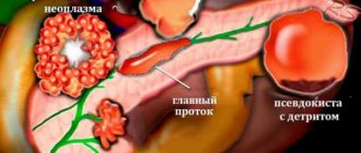
Complications of chronic pancreatitis
- pancreatic pseudocysts;
- biliary obstruction;
- formation of pseudoaneurysms with the development of bleeding;
- thrombosis of the splenic vein.
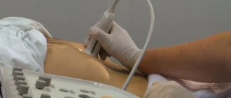
Why is an abdominal ultrasound performed for chronic pancreatitis?
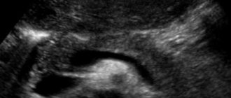
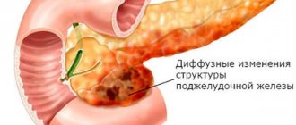
- The parenchyma, inhomogeneous in the early phases, later becomes increasingly atrophic
- The pancreatic duct is dilated, and the presence of stones is often noted.
Chronic pancreatitis. Ultrasound. Dilated pancreatic duct with a stone giving an acoustic shadow. The hyperechoic pancreatic parenchyma is atrophic and narrowed.
What will CT images of the abdominal cavity show in chronic pancreatitis?
- In early forms it is not very informative;
- Initially and during acute inflammation, a circular or diffuse enlargement of the pancreas is detected;
- Later, atrophy of the parenchyma, dilation of the ducts and calcifications are revealed;
- Pseudocysts.
What will RCP show in chronic pancreatitis?
- For moderate and severe forms, the Cambridge classification proposed for ERCP can be applied;
- After the administration of contrast, its heterogeneous accumulation in the parenchyma is observed;
- Intravenous administration of secretin or oral SSGS allows semiquantitative assessment of exocrine pancreatic function.
Is an MRI of the abdominal cavity performed for chronic pancreatitis?
- T1-weighted images show reduced signal intensity from the parenchyma with reduced contrast enhancement due to fibrosis;
- Calcifications are poorly visible;
- On T2-weighted images of areas of acute inflammation, necrotic areas and pseudocysts are hyperintense;
- Dilated ducts are well visualized on T2-weighted images and MRCP.
Chronic pancreatitis. T1-weighted MR image after contrast injection. Large pancreatic pseudocyst with an artery projecting into the lumen of the cyst (risk of bleeding due to arrosion of the vessel)
Why is an abdominal x-ray performed for chronic pancreatitis?
- Calcifications in the projection of the pancreas;
- Currently not used for diagnostic purposes.
How is an MRI of the pancreas performed?
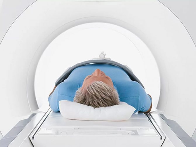
Patient position during MRI of the pancreas
By appointment, you must come to the diagnostic center at the agreed time. The technician places the patient on the tomograph table and secures it with bolsters and straps to ensure maximum immobility. The procedure is monitored from the next room, and there is a button at hand to contact the staff. To improve visualization and obtain more informative images (especially important if a pancreatic tumor is suspected), MRI is performed with contrast: during the study, a paramagnetic drug based on gadolinium chelates is injected intravenously. On the obtained tomograms after enhancement, the structures of the entire biliary system, the size of the neoplasm, and its relationship with surrounding tissues are more clearly visible. The table slides into the lumen of the apparatus ring; many detectors rotate around the desired area inside the tomograph drum, which capture the response of structures to the influence of the magnetic field and transmit the results to the computer. After processing with a special program, three-dimensional images are obtained. A slice thickness of 1.5 mm allows you to see the smallest pathological changes. Pictures can be transferred to any electronic medium or printed. There are no painful sensations, but some patients develop vegetative reactions to the administration of contrast: a metallic taste, a diffuse feeling of heat, nausea, increased salivation, and headache. These symptoms do not require cancellation of the procedure and are considered normal. After a few minutes, after adaptation, unwanted side effects stop on their own. The duration of the study is 45-60 minutes, the wait for the result is no more than an hour. In unclear situations, it is possible to obtain a “second opinion,” which increases the accuracy of diagnosis. Before taking an MRI of the abdominal cavity, pay attention to the technical characteristics of the tomograph: you can get clear images with a power of 1.5 Tesla.
What diseases have symptoms similar to chronic pancreatitis?
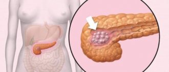
Pancreas cancer
— Atrophy of the parenchyma and dilatation of the pancreatic duct distal to the tumor;
— Signs of tumor infiltration;
— Differential diagnosis may be difficult in the presence of areas of acute inflammation in chronic pancreatitis
Intraductal papillary mucosal neoplasm
— No data on alcoholism
- Usually no calcifications
— Intraductal polyps provide contrast enhancement
Acute pancreatitis
— Severe parapancreatic effusion
— No pancreatic atrophy
How does the technique work?
The accuracy of diagnostics using magnetic resonance imaging reaches 97 percent. The essence of the technique is to monitor contrast, which indicates possible changes.
What does an MRI of the pancreas show?
The magnetic resonance imaging method allows you to determine the size and shape of the pancreas, tissue density, the possible presence of pathological formations (including stones), as well as the patency of the ducts. In particular, using MRI it is also possible to establish:
- localization inside the pancreas;
- dimensions of the organ components;
- fiber condition;
- sizes and types of tumors (if any);
- condition of the capillaries inside the gland;
- presence of metastases.
A highly sensitive device allows you to distinguish benign neoplasms from malignant ones, as well as determine the stage of development of the disease.
Which tomograph is better to undergo the examination?
There are two main types of research apparatus: open and closed. To detect pathological changes in the pancreas, a closed tomograph with a power of no more than one and a half Tesla is used.
Such equipment is highly accurate and can detect even minor anomalies, so it should be chosen for diagnostics.
Devices with a power of less than 1 Tesla are not used for diagnosing the pancreas. In this case, this type is ineffective.
How is the procedure done?
To accurately identify the condition of the pancreas, specialists resort to MRI with contrast. A contrast agent is injected into the patient's vein. It is necessary for coloring organ tissues in a certain color, depending on their condition and density. This leads to a clearer, more contrasty image and allows you to more accurately determine the condition of the gland. Next, the patient lies down on a mobile table and assumes a stationary position. The table is rolled into a tomograph machine and a scan is performed. An MRI can take up to an hour.
Preparing for the MRI procedure
The diagnostic device is highly sensitive and does not tolerate serious errors. Therefore, before the procedures, painless preparation for MRI of the pancreas is necessary, which does not require fundamental changes in the patient’s usual lifestyle.
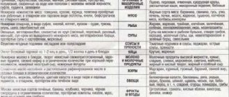
So, first of all, the patient is prescribed a special diet that eliminates the possibility of a large amount of gas accumulation in the intestines. Typically, it begins three days before visiting the doctor's office. Gentle nutrition excludes:
- alcoholic drinks;
- any legumes;
- strong tea and coffee;
- fast food, salted, smoked and fried;
- carbonated drinks;
- baked goods and confectionery products;
- some grain crops;
- fresh milk;
- cabbage and root vegetables.
Depending on the individual characteristics of the patient, other foods may be excluded. Medicines that reduce gas formation can also help you prepare.
Preparation for the examination is not limited to diet. So, other measures need to be taken. To do this, the patient needs:
- temporarily stop taking medications (on an individual basis);
- refuse to eat on the day of the MRI;
- notify the doctor about the possible presence of tattoos, prostheses and other foreign bodies in the body;
- remove all jewelry from the body (if any);
- wear clothes and underwear made exclusively from natural fabrics without hard inserts;
- report any allergies.
When keeping a patient in a hospital, a cleansing enema can be performed, which will help prepare in the shortest possible time. It is important that one or two days before diagnosis the patient does not undergo any additional procedures requiring the administration of contrast agents.
Sequence
MRI diagnostics of the liver and pancreas is carried out in a specially equipped room with a tomograph.
The patient needs to lie down on a specialized diagnostic table, which is then pushed into the labyrinth of the tomograph.
If there is a need to use a contrast agent, then it is administered to the patient intravenously, by installing a catheter connected to a special device that supplies contrast under a certain pressure. In most cases, a contrast agent based on gadolinium is used.
The following drugs used for MRI of the pancreas are most effective:
- Magnevista,
- Primovista,
- Gadovista,
- Prohensa.
All these substances contain various gadolinium compounds. Other iodine-based products are not suitable for MRI. Such drugs are designed for multislice computed tomography, which is carried out on the basis of x-ray examination. Multislice tomography, or MSCT of the pancreas, is prescribed if there are certain contraindications to MRI.
We recommend reading: How cholera is transmitted: the main routes of transmission of the disease
Contraindications to the procedure
Relative contraindications for examination of the pancreas using MRI:
- Contraindications for a neurologist.
- Claustrophobia. For such pathologies, it is preferable to use tomographs with an open type of device.
- Obesity of 2-3 degrees, since standard tomographs are designed for weight up to 120 kilograms.
- Diabetes mellitus using an insulin pump.
- Pregnancy (1st and 3rd trimester).
- Heart failure.
- The presence of a pacemaker in the body.
- Tattoos with added metal, piercings in any part of the body.
- The patient's serious condition.
In all cases, contraindications must be discussed with a doctor; only the doctor can decide on admission to the MRI procedure.
- Previous injuries during which treatment was carried out using the Elizarov apparatus.
- The presence of metal inclusions in the body (prostheses, pins, fragments).
- Stents in coronary vessels.
- Presence of a previous generation pacemaker.
- Clips in the skull.
- Built-in hearing aids.
We recommend reading: Raspberry roots: medicinal properties and contraindications, scope of application and possible harm
Contraindications
Magnetic resonance imaging is considered a harmful procedure and is prescribed with a long periodization. It is not carried out if there are any metal bodies or mechanisms in the body, including:
- Ilizarov apparatus;
- pacemaker;
- clips and implants in the skull area;
- fixed metal crowns and prostheses.
Among the relative contraindications:
- the presence of mental disorders (can be treated with appropriate medications);
- increased body weight (more than two hundred kilograms);
- presence of an insulin pump;
- pregnancy.
As a rule, procedures are not performed if the patient is in a critical or serious condition.