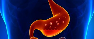
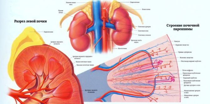
The kidneys are a paired organ that performs an excretory function. They are necessary for removing substances from the human body that remain after metabolic processes. What does such a term as renal parenchyma mean? This is tissue that is located on the surface of this organ and completely covers it. The main functions of the parenchyma include maintaining the process of formation and excretion of urine.
If for some reason the structure of this tissue is disrupted, serious changes occur in the human body. This condition must be promptly identified and eliminated, which will avoid many negative consequences.
General information about parenchyma
Parenchyma is the tissue that completely covers the outside of the kidneys. In turn, this structure consists of two more layers - cortical and medullary. It is very thin and is a collection of small capsules that are well supplied with blood. It is these elements that are responsible for the production of urinary fluid. If you look at both organs, then you can find more than a million of these capsules. The fluid they produce leaves the parenchyma and collects in the pelvis and calyces.
A special feature of this tissue structure is that it is capable of changing in a certain way throughout a person’s life. In children, its thickness is 13-16 mm. During puberty (after approximately 16 years), the parenchyma becomes thinner. Its thickness reaches 11-13 mm. Subsequently, the structure and size of this layer of buds does not change. If any deviations from the norm are detected, the presence of certain diseases that require special treatment can be suspected.
Main indicators of parenchyma: norm and pathology
Thickness
The normal thickness of the kidney parenchyma is 15-25 mm. In elderly patients over 60 years of age, this figure is slightly lower - about 11 mm. If it deviates either upward or downward, it is necessary to promptly establish the cause.
To understand, you need to remember the anatomy
Table: Reasons for deviation from the norm:
| Thickening of the renal parenchyma | Thinning of the kidney parenchyma |
| Acute inflammatory processes (for example, with glomerulonephritis) | Chronic glomerulonephritis |
| Acute nephrotic syndrome | Infectious diseases |
| surge arrester | Chronic inflammatory lesions of the parenchyma |
| Cysts | Neoplasms originating from epithelial tissue |
| Tumors and other neoplasms originating from parenchymal tissue | Hydronephrosis |
| Compensatory (vicarious) kidney hypertrophy |
If we compare all parenchymal dystrophies, the most common is a decrease in the kidney parenchyma. This condition is fraught with a decrease in the number of functioning nephrons and the development of renal failure. The final stage of the pathological process is parenchymal atrophy.
Structure
When a patient undergoes an ultrasound of the kidneys, he may often find in the conclusion an indication that the echostructure of the parenchyma is changed. What does it mean?
Normally, the structure of the organ tissue is homogeneous, without pathological inclusions. With any disease, diffuse or focal changes can be observed.
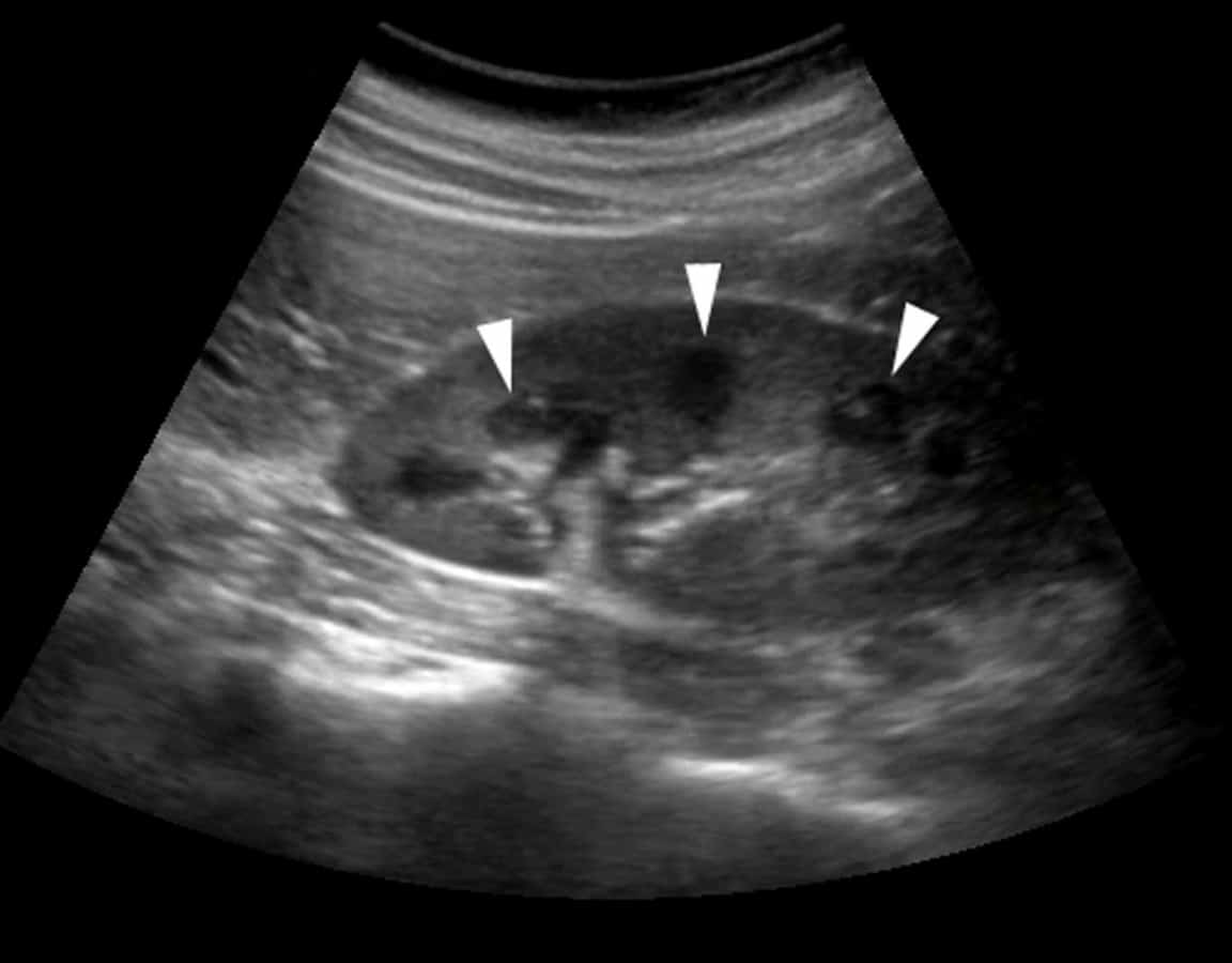
This is what healthy kidneys look like on an ultrasound
Uniformly heterogeneous renal parenchyma is observed with:
- development of urolithiasis;
- inflammatory, including autoimmune diseases;
- some endocrine diseases - diabetes mellitus, hyperthyroidism;
- atherosclerosis of renal vessels.
- diffuse metabolic changes (a typical example is calcifications in the parenchyma).
Focal
Limited (focal) changes in the parenchyma are observed with:
- benign neoplasms (oncocytoma, angiomyolipoma, adenoma);
- malignant neoplasms (cancer);
- cysts.
A pathological focus can also be caused by a stone in the parenchyma.
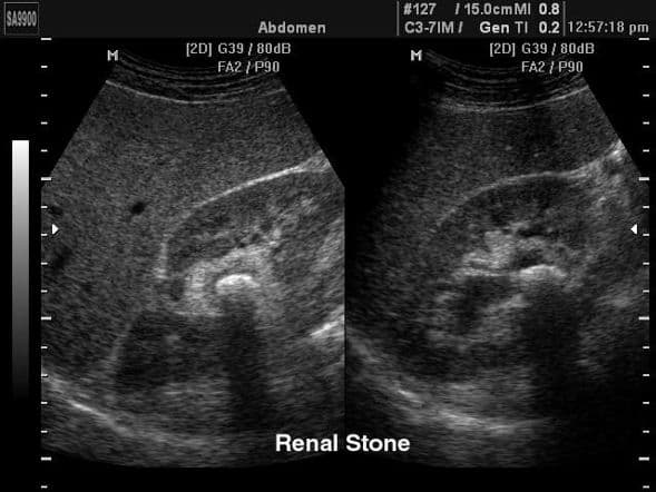
The calculus is well visualized
Density
The echogenicity of the parenchyma is its ability to reflect or transmit ultrasonic waves.
Normal on ultrasound:
- the pyramids have low echogenicity (almost black);
- the cortex and columns are isoechoic and identical to each other (gray);
- sinuses are hyperechoic (the lightest).
Most often in medicine, increased density of the kidney parenchyma is encountered.
This may indicate development:
- diabetic nephropathy;
- chronic infectious diseases;
- consequences of arterial hypertension;
- glomerulonephritis;
- amyloidosis;
- sclerotic changes.
If the echogenicity of the renal parenchyma in the fetus is increased, this is a consequence of congenital developmental anomalies. Severe parenchymal fibrosis should alert the physician.
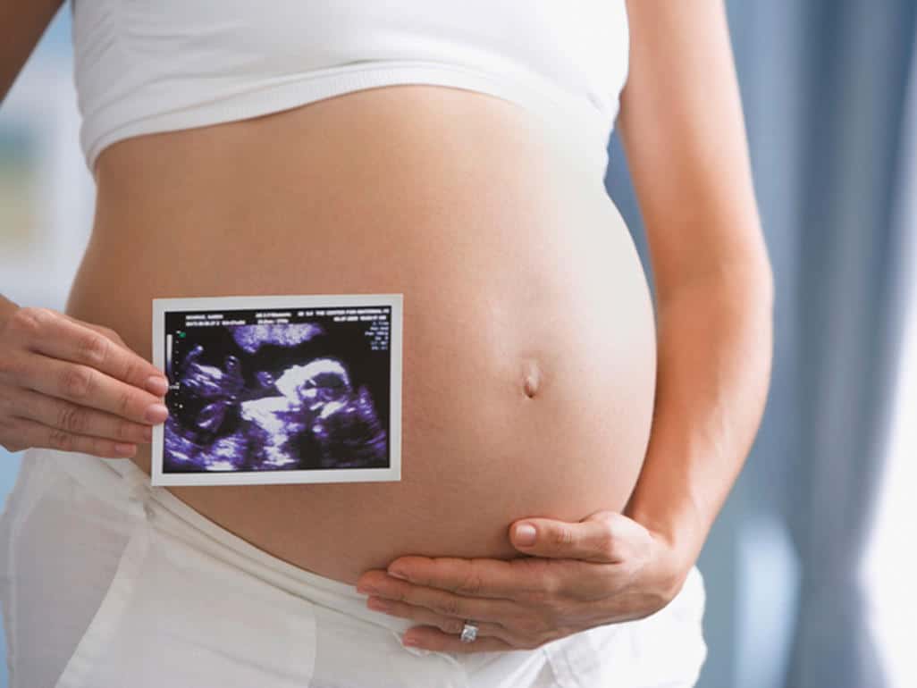
During pregnancy, it is very important to undergo screening ultrasound examinations
In adults, the renal parenchyma is denser more often in chronic diseases, chronic renal failure. Typically, this pathology is diagnosed in elderly patients. As a rule, parenchymal fibrosis is a consequence of the replacement of functioning nephrons with connective tissue.
Note! Ultrasound diagnostic doctors are well acquainted with so-called pseudopathologies. For example, sometimes enlarged Bertin's columns extend quite deeply beyond the parenchyma into the renal sinus. It seems that such a parenchymal bridge of the kidney literally divides the organ in two. However, this is not a tumor or pathological tissue, but simply an individual characteristic of the patient.
Parenchyma cells are an important structural element of any organ. Kidneys are no exception. Regular preventive examinations and disease prevention ensure that they remain healthy for a long time.
Increased echogenicity
If increased echogenicity is detected during ultrasound, this may indicate the following:
- development of the inflammatory process;
- the presence of endocrine problems;
- developing glomerulonephritis.
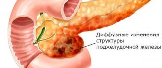
Diffuse changes - causes and consequences
Diffuse changes in the parenchyma can occur in several directions:
- thickening or thinning of the tissue layer;
- the appearance of areas characterized by increased or decreased echogenicity;
- change in the nature of arterial blood flow in the upper layer of the kidney;
- the appearance of pathological inclusions containing liquid;
- change in the size of the kidneys relative to each other.
When identifying such pathologies, it is necessary to conduct a serious examination of the body, since this indicates the presence of chronic diseases of the urinary system. If nothing is done, the underlying parenchyma will begin to thin out, which is very dangerous.
These disorders most often occur against the background of the following diseases:
- the development of urolithiasis, which negatively affects the structure of the kidneys;
- the presence of inflammatory processes in various tissues. Most often they occur in the nodules or canals of the kidneys;
- various kinds of endocrine disorders. Hyperthyroidism, diabetes and other diseases can lead to such negative consequences.
Reduced thickness of the upper layer of the kidneys
If an ultrasound examination reveals that the tissue covering the kidneys is thinned, serious health problems may be suspected. Most often, this disorder is detected during the course of certain infectious diseases or while taking certain medications. Slight thinning also occurs with age.
This pathological condition develops gradually, so a person discovers the problem when it has worsened significantly. With a critical decrease in the thickness of the parenchyma, it ceases to function normally. This is extremely dangerous, therefore, if such a problem is detected, treatment and constant monitoring of indicators must occur.
Pathological changes
Kidneys normally have a number of characteristics, including certain sizes:
- length up to 10–12 cm;
- width within 5–6 cm;
- thickness up to 4 cm;
- the total weight of one kidney is up to 200 g.
One of the negative types of tissue structure transformations are diffuse changes in the kidney parenchyma. They indicate an enlargement of the organ and may indicate the following pathologies:
- congenital;
- age;
- chronic;
- acquired as a result of any infections.
The causes of diffuse changes lie in a number of diseases:
- urolithiasis disease;
- urinary tract lesions;
- inflammatory processes in the tubules, which form the inner part of the parenchymal tissues, and the vascular system of the kidneys;
- fatty plaques in the pyramids, which are located in the medulla.
Calcifications
Kidney stones very often lead to changes in their structure. This problem is caused by poor nutrition and the presence of certain diseases in which metabolic processes in the body are disrupted. Only calicinates can form in the parenchyma. They form on the surface of dead tissue. Calcium salts are deposited on them, which eventually leads to the formation of stones.
This problem can occur at almost any age in a person, both female and male. In the presence of calcifications, edema very often develops. Treatment of this disease can cause some difficulties. Therefore, it is easier to follow some rules to prevent the formation of kidney stones. These include following healthy eating recommendations and timely treatment of any inflammatory diseases. You should also not limit physical activity and play sports all the time.
Diffuse kidney changes
Diffuse changes are a very broad spectrum concept. It reflects the final conclusion of the study and is not a diagnosis as such. Signs of chronic organ disorders provoke thinning of the tissue, and infectious effects lead to its thickening and enlargement.
An increase can be provoked by: the presence of fluid in the renal pelvis, a developing abscess, the formation of thrombosis, deformation of the venous structure of the organ or kidney stones.
Signs of such changes may include:
blood in the urine pain when urinating pain in the hypochondrium, during an external examination of the patient
If, after the study, the following were discovered: asymmetry of the shadows of the kidneys, uneven contours, any obvious deformation of the brain area, uneven tissue thickness, then this indicates the progression of pyelonephritis, which requires immediate treatment.
The concept of echogenicity is one of the main ultrasound parameters, which reflects the degree of tissue density. Echogenicity decreases as a result of some kind of inflammatory process or swelling.
Thus, the less liquid the structure under study has, the higher its echogenicity will be; the more liquid, the lower this parameter.
Increased echogenicity is a consequence of the following diseases and abnormalities:
diabetic nephropathy glomerulonephritis metabolic disorder inflammation
Normally, the tissue on a kidney ultrasound will be homogeneous.
The appearance of cysts
If the nephrons retain fluid, cysts can form in the top layer of the kidneys. Their number can be almost any – from one to several pieces. Cysts are formations with thin walls that are filled with fluid. The danger of this disease is that it can affect the sinuses of the kidneys.
If cysts are detected, it is recommended to remove them surgically. After such manipulations, the inflammatory process disappears, and the functioning of the organ returns to normal. The critical diameter of cysts, which does not pose a serious danger to humans, is considered to be 10 mm or less.
Symptoms of this pathology include the presence of intense pain in fasting, bloody inclusions in the urine and high blood pressure.
Symptoms of structural changes
A visit to a urologist or nephrologist is required if negative signs appear:
- pain in the lumbar region;
- deterioration in the quality of urine: the appearance of mucus, blood, “flakes”, sediment, cloudy urine;
- painful pain when emptying the bladder;
- swelling of the eyelids, legs;
- more frequent or rare urination due to normal drinking regimen;
- increased blood pressure in combination with edema.
Based on the results of ultrasound, doctors identify various structural changes in the renal parenchyma:
- accumulation of fluid in the renal pelvis;
- violation of the venous structure;
- thrombosis;
- inflammatory process;
- the presence of sand or kidney stones;
- thickening or thinning of the parenchyma;
- cystic formations;
- tumors (benign or malignant);
- increase in kidney size;
- disturbance of arterial blood flow;
- areas with reduced or increased echogenicity.
Learn about the symptoms and treatment of kidney stones with stone dissolving tablets.
Effective methods of treating acute pyelonephritis in women are described on this page.
Follow the link and read about the rules and features of nutrition for kidney failure.
Presence of tumors
Both benign and malignant tumors can form in the parenchyma. The first type can always degenerate into oncology. Therefore, if any tumors are detected, a thorough examination (ultrasound, CT) is indicated, after which removal surgery is performed.
The sooner you take the necessary measures, the higher the likelihood of a positive result. You should not neglect the recommendations of doctors, as this is dangerous to health and life.
A secret from our reader
Have you ever suffered from problems due to CYSTITIS? Judging by the fact that you are reading this article, victory was not on your side. And of course you know firsthand what it is:
- At the initial stage of the disease, the lesion is observed only on one foot. Later it goes to the other side.
- Increased frequency of urination up to several times per hour.
- Reducing the amount of urine.
- Soreness above the pubis and behind the pubis.
Parenchyma cells, as a rule, have rounded outlines, however, they can also be elongated. In plants, water and minerals move through the walls of such cells. In different parts of the plant, the parenchyma can change and acquire specialized properties. These cells include the epidermis, a thin covering tissue. It consists of a single layer of cells and covers the entire primary body of the plant. The main function of the epidermis is to protect plants from drying out and from penetration of pathogenic organisms.
Assimilation parenchyma is a specialized tissue containing a large number of chloroplasts (chlorophyll-bearing cells of the leaf, stem, bark). Its main function is to carry out the processes of photosynthesis. Plant parenchyma cells provide support for the organs in which they are located. This property is especially important for the stems of herbaceous plants. Unspecialized parenchyma cells remain metabolically active; many processes important for the plant organism take place in them. Through a system of intercellular spaces filled with air, gas exchange occurs between the external environment and living cells. Parenchyma cells also serve as nutrient storage.
What are the dangers of changes in parenchyma?
The doctor can judge the condition of the kidneys . Changes in the parenchyma indicate an inflammatory process in the kidneys, which developed as a result of untimely treatment of renal disease.
Thinning
We can talk about thinning of the parenchyma if its thickness is less than 1 cm.
This indicates serious renal pathologies with a long-term chronic course . If the disease proceeds slowly, then the parenchyma becomes thinner gradually. During an exacerbation, thinning occurs rapidly and the organ may lose its functions, which poses a direct threat to life.
The main causes of thinning:
- kidney infections;
- viral diseases (flu);
- kidney inflammation;
- inappropriate treatment of kidney diseases.
Thickening
An increase in the size of the parenchyma is also a symptom of serious kidney damage . Among these diseases:
- kidney cysts;
- malignant neoplasms of organs;
- kidney tuberculosis;
- inflammatory diseases.
With any pathological change in the parenchyma, the basic function of the kidneys is disrupted. They are no longer able to remove harmful substances from the body. The patient shows signs of intoxication :
- temperature increase;
- pain when urinating;
- swelling of the legs and arms;
- cloudiness of urine, change in color.
If one kidney is affected, the second compensates for the impairment, taking over all functions. The greatest danger is damage to both kidneys . If the disease is neglected, the kidneys will never be able to function normally. The only chance to prolong life is regular hemodialysis or a kidney transplant.
Tumors
Thickening of the parenchyma is dangerous because it increases the risk of formation of growths in the kidneys . According to statistics, most growths are malignant in nature. The main symptoms of kidney cancer are:
- sudden weight loss;
- phlebeurysm;
- increased blood pressure;
- sudden temperature changes.
If cancer is detected at an early stage, surgery is performed to remove the tumor or the entire kidney. This increases the patient's likelihood of survival .
Another common cause of thickening of the parenchyma is cystic growths. They are formed due to fluid retention in the nephrons. Typically, such cysts are up to 10 cm in size. After removal of the cyst, the renal parenchyma acquires normal thickness.
Echogenicity
Also an alarming symptom is an increase in the echogenicity of the kidney . This condition is determined using ultrasound. Increased echogenicity indicates diseases such as:
- diabetic nephropathy;
- glomerulonephritis;
- urolithiasis disease;
- endocrine system disorders;
- extensive inflammatory processes.
Parenchyma in the human body
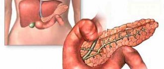
The parenchyma also plays an important role in the human body. It is the main functional tissue of parenchymal organs: liver, spleen, lungs, kidneys, pancreas and thyroid gland. It consists of connective tissue stroma and specialized cellular elements. Parenchyma can be formed by various types of tissue: epithelium (glands), hematopoietic tissue (spleen), nerve cells (nerve ganglia). The lung parenchyma is part of the apparatus that carries out external respiration. It consists of pulmonary acini. The pulmonary acini begin with a terminal bronchiole, which branches successively into respiratory bronchioles, alveolar ducts, and alveolar sacs, making up the alveolar tree. External respiration occurs in the lung parenchyma, one of the elements of which is the diffuse exchange of gases.
Kidney parenchyma cells are a specific tissue that performs the main function of this organ. The spleen is also a parenchymal organ. Its parenchyma is a collection of lymphoid cells. Another organ, the liver, consists entirely of parenchymal tissue, which is made up of hepatocytes. The pancreatic parenchyma is a multi-structured tissue, which consists of numerous irregularly shaped lobules and round cellular areas (islets of Langerhans). Diseases of the parenchyma include numerous benign and malignant neoplasms with different structures. Among them, renal parenchyma cancer is quite common, accounting for about 90% of all cases of tumors of this tissue.
How the organ works
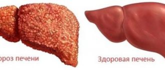
The liver tissue is divided into special lobules, between which blood and bile ducts are laid. This structure, created by nature, is ideal, since it provides both blood access to literally every cell of the organ and the removal of substances synthesized by the cell. During a person’s life, negative changes in the liver parenchyma occur to a greater or lesser extent; this practically cannot be avoided.
Poor diet, alcohol, toxic medications, smoking, polluted environment, sedentary lifestyle - all this, as well as many other factors, cannot but affect our liver, disrupting its functions.
Negative transformations of the liver parenchyma can occur as a result of exposure to viruses and toxins, metabolic disorders, in particular lipid, etc. As a result, various diseases arise, such as hepatitis of various etiologies, fatty liver, replacement of functional cells (hepatocytes) with fibrous tissue and etc. In especially severe cases, liver failure and cirrhosis may develop.
Unfortunately, pathological changes in the liver tissue are not always possible to identify immediately. In any case, traditional ultrasound examination is practically powerless here. Only a comprehensive and comprehensive examination of the entire gastrointestinal tract can tell a specialist that the patient has begun to experience such changes.
Note to the patient
At home, it is impossible to identify the disease that provoked the development of diffuse changes. In such cases, an ultrasound is prescribed. If echo signs of diffuse changes in the renal parenchyma appear, it is recommended to seek help from a doctor. First of all, the specialist will prescribe a complete examination of the patient. Based on the results of the study, he will prescribe a comprehensive treatment, during which it is necessary to lead a healthy lifestyle and not engage in heavy physical labor, eat right, and avoid stress and anxiety.
There are several diseases that can trigger the occurrence of such a pathology. At the initial stage of development of the disease, treatment is carried out more effectively with the help of medications. In advanced cases, surgical intervention is necessary. Under such circumstances, the patient recovers significantly longer. To reduce the risk of pathology, experts recommend regular medical examinations, since there are cases when at the initial stage of development of diffuse changes in the parenchyma there are no symptoms. If someone in your family has had a similar disease, you should visit the hospital once every six months.
Source: fb.ru
Source: gepasoft.ru
What are the changes in organ tissue?
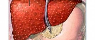
Pathological changes in the liver can be of two types:
- Focal;
- Diffuse.
As you might guess, focal changes are those that do not involve the entire parenchyma, but only individual ones, and this is the coverage of the entire liver tissue by the pathological process.
As a simple example of focal and diffuse changes in liver tissue, the following information can be given:
- Hepatitis or cirrhosis are diseases that cause damage to the entire parenchyma;
- , abscess, malignant tumor of the liver - ailments that affect the parenchyma focally, without spreading to its entire surface.
For example, a diffuse form of cancer affects the entire organ, practically making it impossible for the doctor to detect unchanged tissue. Or variants in which, against the background of diffuse changes, there are also focal ones, for example, an abscess against the background of ongoing hepatitis.
Separately, it should be noted that in a number of cardiovascular pathologies, tissue edema usually develops. The liver parenchyma is no exception here. Doctors usually refer to this pathology with the term “”. At the very beginning of the development of edema, the parenchyma usually looks homogeneous, but as the condition worsens, fibrous foci gradually develop, forming heterogeneity of the liver tissue. In particularly severe cases, fluid begins to accumulate in the abdominal cavity and ascites develops.
Regenerative capacity of tissue
To the great happiness of mankind, wise nature has provided that pathologically altered liver tissues are in most cases capable of regeneration.
It turns out that our multifunctional vital organ has colossal internal potential, sometimes being recreated even after the loss of almost three-quarters of actively functioning cells!
If the liver did not have this ability, humanity would be practically unviable.
However, in many cases the body becomes impossible, and then a transplant surgeon comes to the patient’s aid. Patients who have irreversible damage and there is no possibility of effective conservative therapy undergo a donor organ transplant.
In recent years, attempts have been made around the world to create a so-called bioengineered liver, which can replace a natural organ, at least temporarily. The bioframework of the future organ has already been created, and the next task, more complex, is the synthesis of liver parenchyma.
Large medical dictionary Atlas of human anatomy Psychological encyclopedia Dictionary of analytical psychology Explanatory dictionary of psychiatric terms Dictionary of neurolinguistic programming Dictionary of medications Biological encyclopedia Dictionary of microbiology Agricultural encyclopedic dictionary Veterinary encyclopedic dictionary Anatomy and morphology of plants Life and fishing of freshwater fish Animals of Russia. Directory of Breeds of Farm Animals. Directory Terms and definitions used in animal breeding and genetics Philosophical encyclopedia History of philosophy Chinese philosophy. Encyclopedic Dictionary. Dictionary of Logic Terms Terms of Gender Studies Brockhaus Biblical Encyclopedia Concise Church Slavonic Dictionary Islam. Encyclopedic Dictionary. Buddhism Religious terms Dictionary of oriental terms Musical encyclopedia Russian rock. Small Encyclopedia Large Encyclopedia of Culinary Arts Encyclopedic Dictionary F.A. Brockhaus and I.A. Ephron Modern Encyclopedia Big Encyclopedic Dictionary Chemical Encyclopedia Natural History. Encyclopedic dictionary Astronomical dictionary Human ecology Rules of Russian spelling Dictionary of management Grammatological dictionary Explanatory dictionary of Ozhegov Modern explanatory dictionary of the Russian language Efremova Explanatory dictionary of Dmitriev Stylistic encyclopedic dictionary of the Russian language Russian spelling dictionary Five-language dictionary of linguistic terms Etymological dictionary of the Russian language by Max Vasmer Etymological dictionary of the Russian language Semenova Etymological Dictionary Russian language Dictionary of the Old Russian language (XI-XIV centuries) Michelson's large explanatory and phraseological dictionary (original spelling) Educational phraseological dictionary Dictionary of popular words and expressions Large dictionary of Russian sayings Explanatory translation dictionary Living speech. Dictionary of colloquial expressions Russian verbal stress Dictionary of Russian idioms Dictionary of antonyms Dictionary of literary terms Poetic dictionary Bulgakov's Encyclopedia Shakespeare's Encyclopedia Dictionary of scribes and bookishness of Ancient Rus' Proper name in Russian poetry of the 20th century: dictionary of personal names Ancient writers Encyclopedia of cultural studies Dictionary of medieval culture Postmodernism. Dictionary of terms Large philatelic dictionary Dictionary of GOST vocabulary Counterintelligence dictionary Armed forces of foreign countries Encyclopedia of fashion and clothing Commercial electric power industry. Dictionary-reference book Large encyclopedic polytechnic dictionary Scientific and technical encyclopedic dictionary Dictionary of metallurgical terms Marine dictionary Technical railway dictionary Encyclopedia of medieval weapons Geographical encyclopedia Demographic encyclopedic dictionary Cities of Russia St. Petersburg (encyclopedia) All of Japan Architectural dictionary Geological terms Dictionary of gold mining Russian Empire Ecological Dictionary Dictionary measures Legal Dictionary Economic Dictionary Encyclopedic Dictionary of Economics and Law Large Legal Dictionary Administrative Law. Dictionary-reference book Dictionary of depository terms Diplomatic dictionary Dictionary of personal names Historical dictionary 1000 biographies Encyclopedic dictionary of pseudonyms Encyclopedia of Russian life of the 19th century All monarchs of the world Encyclopedia of battles of world history Ancient world. Dictionary-reference book Encyclopedia of the Third Reich
The human body cannot cope with poisons and toxins without the liver. But when exposed to these substances, the liver parenchyma (its structure) is destroyed. Harmful substances are not restrained by anything and they gradually poison everything in the human body. Therefore, timely detection of pathology of the liver structure and treatment is the key to a healthy liver and the body as a whole.
Bad habits, ecology, and diseases can provoke destruction of the liver structure, which will lead to intoxication of the entire body.
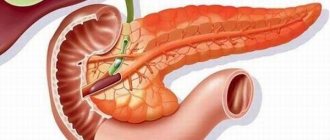
Structure of the liver: structure and functions
The liver is located in the upper right part of the abdomen under the ribs. The upper border reaches the level of the nipples. Parenchyma is the tissue that forms the liver. The parenchyma consists of hepatic lobules. The lobes have a prismatic shape and fit into each other. Between them there is an intermediate substance, blood vessels and bile ducts. This structure is optimal for the human body, since each cell of the organ receives a sufficient amount of blood and each of them has a outlet for synthesized substances. Each lobe is from 0.7 to 2 mm in diameter. There are about 1 million such components of the parenchyma.
The liver performs the following functions in the body:
- secretory - the main function is the secretion of bile and its transportation through the bile ducts;
- endocrine - synthesis and secretion of albumin, globulin, fibrinogen, lipoproteins, prothrombin, glucose and other substances;
- metabolic - normalizes the metabolic processes of proteins, amino acids, fats, carbohydrates, vitamins and hormones.
But besides this, the liver processes and absorbs drugs and steroids. It is responsible for maintaining normal blood sugar levels. In addition, it accumulates useful substances from metabolic products. One of the most valuable properties of the liver is the neutralization of poisons.
Special Kupffer cells are responsible for this, which bind harmful substances and remove them into the intestines.
Types of changes
There are several types of changes in the liver parenchyma: in its composition, shape or density. The extent to which these changes are expressed and their essence is determined by one of the types:
- focal - single damage or compaction;
- local - a separate homogeneous part of the parenchyma is damaged;
- diffuse - damage to the parenchyma as a whole.
Focal type
Focal changes are diagnosed using echography. The degree of echo reflection divides focal lesions into those without echostructure, those with weak, strong and mixed echostructure. The lesions can be single, multiple or merging. If the patient's condition worsens, the echogenicity of the lesion may change. Also, echography reveals the appearance of single or multiple calcifications (highly echogenic compactions) in the parenchyma. Most often observed in adults, they appear rarely in children. Occurs in patients with the following concomitant diseases:
A focal lesion devoid of echostructure is a parenchymal cyst. During echography they appear if their diameter is 3-5 mm, because only with this size do they have sufficient echogenicity. There are several types of cystic neoplasms, they are distinguished depending on:
Diffuse changes are hepatitis (in acute or chronic form), fat accumulation, cirrhosis, structural changes under the influence of other diseases. In a patient with hepatitis, the liver becomes larger, but the parenchyma remains the same as it was. But if the inflammation intensifies, the parenchymal surface will change, causing the thin liver wall to increase in size. When echography, decreased echogenicity and increased sound conductivity are observed. With hepatitis, non-homogeneous inflammation of the parenchyma leads to the fact that echo density can be high in one area and low in another.
With cirrhosis, the number of affected areas with impaired echogenicity increases significantly, because the homogeneous structure is destroyed much faster. Echo density is significantly reduced. The sizes of such areas range from 0.5 cm to 2 cm. Such a heterogeneous structure of the liver parenchyma can also be caused by stagnation in the bile ducts, fatty degeneration of the parenchyma, and impaired metabolism.
Definition of the word “Parenchyma” according to TSB:
Parenchyma (from the Greek parеnchyma, literally - poured nearby) 1) the main tissue of plants, consists of cells of more or less the same size in all directions. P. cells form homogeneous clusters in the plant body, fill the spaces between other tissues, and are part of conductive and mechanical tissues. Due to the functional specialization of protoplasts, P. cells can perform assimilation, excretion, and other functions. The presence of intercellular spaces in P. (especially loose, see Fig.) determines its participation in gas exchange. Parenchyma cells that perform a supporting function can be elongated, branched, stellate (sclereids); they have thick, often lignified walls. Living parenchyma cells are capable of dividing. Phelogen (cork cambium) is formed in the plant, and in plants with an atypical increase in thickness, the cambium is formed (beet roots, some vines). 2) In animals, P. is a phylogenetic predecessor of real tissue. They are distinguished: primary P. - a connection of a large number of homogeneous cells located without a specific system, not fused into a syncytium and not separated by an interstitial substance (for example, in the embryos of some hydroids at the morula stage), and mixed P. - a connection of heterogeneous cells located without a specific order (for example, in the body of intestinal turbellarians). Sometimes P. is called the main functional tissue of the liver, spleen, lung, gland, or striated muscle. Cross section of the stem of the spreading rush: a - epidermis. b — assimilation parenchyma. c - conducting bundles. d - loose parenchyma of the core.
Signs and symptoms
The most common symptoms of damage to the liver parenchyma:
- headache;
- feeling of nausea;
- bitter taste in the mouth;
- pain under the ribs on the right side;
- yellow skin tone;
- weakness in the body.
If a person has any of the listed complaints, then he should consult a doctor in order to identify the cause in time and begin treatment. The first study that is performed is an ultrasound. As a result, various types of damage can be detected in the homogeneous structure of the parenchyma. Diffuse damage in small quantities may indicate recent viral diseases or poor nutrition; they are not harmful to human health. But if the changes are significant, this indicates that they are caused by serious diseases. Therefore, additional laboratory tests should be performed.
It is necessary to know what signs of changes in the structure of the liver exist. Jaundice caused by parenchymal disorders manifests itself as follows:
- the sclera turns yellow, followed by the mucous membrane of the palate and skin (may have red and green shades);
- the skin becomes inflamed and itchy;
- feces become discolored and urine darkens;
- body temperature rises;
- the person feels sick and vomits;
- muscles and joints hurt.
Focal inflammation of the liver also has characteristic signs:
- calcifications in the liver (hardening consisting of calcium salts)
- liver cysts.
Causes of hepatic parenchyma
Viruses, alcohol, and complications of other diseases cause pathological modifications of liver cells.
Since the main symptom is yellowing of the skin, it is necessary to understand how it occurs. It is caused by improper functioning of liver cells (hepatocytes). They are responsible for binding bilirubin to glucuronic acid (converting it from indirect to direct). If the liver is damaged or inflamed, its cells are affected and, as a result of the damage, are unable to take up bilirubin. Due to the accumulation of this pigment in the blood, jaundice occurs. Another reason for this symptom is stagnation of bile.
There are a number of root causes for the development of liver pathology:
- infection with viruses (viral hepatitis);
- poisoning by toxins (at home, in industrial production, during treatment) and alcohol;
- sepsis, which causes a lack of oxygen in cells;
- autoimmune type of hepatitis (the immune system attacks the liver parenchyma with antibodies).
Ultrasonography
The advantage of ultrasound is that it does not require special preparation for the procedure. The patient is required only 3 days before the study not to eat foods that provoke increased formation of gases (legumes, white cabbage, soda, dark bread, grapes). It is recommended to perform an ultrasound on an empty stomach, so the results will most accurately reflect the real picture of the disease. But if it is contraindicated for a patient to skip meals, as, for example, with diabetes, then you should not deviate from the doctor’s instructions.
Normal picture of parenchyma on ultrasound
The normality of the parenchyma is assessed by the size of the hepatic lobes, the delineation of their contours and structure.
These parameters are compared with the norm. This way the doctor will know which one is affected. If a person has such dimensions, then his liver is healthy:
- the size of the right lobe is no more than 12.5 cm;
- the size of the left lobe is no more than 7 cm;
- diameter of the portal vein - no more than 13 mm;
- common bile duct - no more than 6−8 mm;
- The edges of the liver are smooth.
Sonographic signs of pathologies
Pathology may be indicated by increased echogenicity and heterogeneous echo density. Such signs imply serious liver diseases: cirrhosis, hepatitis, fatty hepatosis. In this case, a biopsy of the defective areas of the parenchyma is necessary. Only in this way will the doctor be able to accurately determine the cause of the problem. If manifestations of increased echogenicity are combined with focal inflammation, this indicates an abscess, hematoma or metastases in the liver. Such processes sometimes indicate lymphoma.
Increased echogenicity of the renal parenchyma - is it dangerous?
According to statistics, today, against the background of general morbidity, people more often suffer from problems of the urinary system. Pathological processes in the kidneys cannot always be observed; more often they occur hidden.
Echogenicity of the kidneys can be diagnosed using ultrasound.
The technique is invasive, is completely painless and has a big advantage : using ultrasound, you can detect the slightest pathological changes even in the early stages.
This will increase the patient's chances of recovery. The diagnostic process itself takes no more than 20-25 minutes, during which time you can find out such parameters as:
the size of the organ itself, its location, neoplasms, if any.
Increased echogenicity of the kidneys may indicate:
diabetic nephropathy (enlarged kidneys, but the pyramids located in the medulla have reduced echogenicity); glomerulonephritis , which occurs in severe form, and the renal parenchyma itself diffusely increases its echogenicity. increased echogenicity of the renal sinus indicates that there are inflammatory processes, metabolic and endocrine disorders .
Kidneys whose tissue is healthy have normal echogenicity; it is homogeneous on ultrasound.
Other diagnostic methods
Modern medicine includes many ways to detect diseases in the liver, thanks to which the most accurate diagnosis can be made.
In order to obtain adequate research results, it is not enough to perform only ultrasound diagnostics and echography on the patient. The examination should be comprehensive and include:
- general tests: blood, urine, feces;
- biochemical blood test;
- ELISA, PCR, which detect antibodies capable of resisting viruses, their DNA or RNA, and thereby confirm their role in the development of the disease.
- tomography (CT);
- liver biopsy with histological examination of a fragment of the affected area is done if cirrhosis is suspected (formation of fibrous nodes in the liver).
Regenerative ability
Regeneration of the liver parenchyma is activated in response to the death of its cells. Regenerating cells contain a large supply of glycogen and normal organelles. The main manifestation of regeneration processes is the accelerated division of liver cells. Due to this, the thin liver surface grows and replaces dead areas. The bile ducts can also be restored. In particularly severe cases, when the liver is irreversibly damaged, the patient requires a transplant.
Treating Changes
Treatment is aimed at identifying the root cause of pathologies of the liver structure and eliminating it. If a patient is diagnosed with a viral disease (hepatitis), he is prescribed antiviral drugs (Viferon, Alfaferon). In case of autoimmune diseases, the patient is supposed to take immunosuppressive drugs (Azathioprine, Prednisolone). It is forbidden to drink alcohol so as not to aggravate the condition of the parenchyma.
In addition to the main treatment, amino acids, phospholipids and vitamins are prescribed. Phospholipids are responsible for the accelerated restoration of cellular structure, amino acids and vitamins are responsible for eliminating the deficiency of nutrients.
In combination with medications, a strict diet is always followed. Spicy, salty, fried and fatty foods are completely excluded from the diet. To help your liver cleanse itself, you should eat more foods high in fiber and potassium. Herbal preparations, decoctions, and infusions also effectively heal the liver. Folk remedies that are used to restore the liver: pumpkin juice with honey, plum juice, decoctions of chicory root, rose hips, oats.