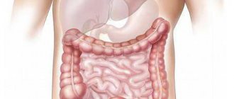
Structure of the small intestine
The intestine is the longest organ of the digestive system and consists of two sections. The small intestine, or small intestine, forms a large number of loops and continues into the large intestine. The human small intestine is approximately 2.6 meters long and is a long, tapering tube. Its diameter decreases from 3-4 cm at the beginning to 2-2.5 cm at the end.
At the junction of the small and large intestines there is an ileocecal valve with a muscular sphincter. It closes the exit from the small intestine and prevents the contents of the large intestine from entering the small intestine. From 4-5 kg of food gruel passing through the small intestine, 200 grams of feces are formed.
The anatomy of the small intestine has a number of features in accordance with its functions. So the inner surface consists of many semicircular folds. Thanks to this, its suction surface increases 3 times.
In the upper part of the small intestine, the folds are higher and located closely to each other; as they move away from the stomach, their height decreases. They may be completely absent in the area of transition to the large intestine.
Where is the ileum located and how does it hurt?
The small intestine ends in the intestine, which due to its location is called the ileum. It is located in the iliac fossa, to the right of the midline of the abdomen.
It has a common mesentery with the jejunum, with which they are attached to the peritoneum, consisting of two layers of peritoneum with a fatty layer between them, penetrated by blood vessels and nerve fibers.
Because of the single mesentery, scientists consider the ileum and jejunum to be one organ. The location of the upper loops of the ileum is vertical, the lower ones are laid horizontally.
Where is the ileum located in humans?
The ileum (lat. ileum) is the lower section of the small intestine, coming after the jejunum. The ileocecal valve separates the cecum from the upper part of the colon.
The ileum is located in the lower right part of the abdominal cavity and in the region of the right iliac fossa. The ileum is covered on all sides by peritoneum. The ileum has a well-defined mesentery.
There is no pronounced anatomical structure separating the jejunum and ileum. At the same time, the ileum has a larger diameter, its wall is thicker, and it is richer in blood vessels.
In relation to the midline, the loops of the ileum lie mainly on the right, the loops of the jejunum on the left.
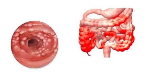
Diseases of the ileum
Intestinal diseases have relatively similar symptoms; most often they are provoked by disturbances in the absorption of substances, the amount of excretion, and digestion. If you have frequent stomach rumbling, upset, frequent and regular pain, you need to undergo examinations in order to diagnose a possible disease in the early stages.
If you ignore the above symptoms, then after 2-4 years the disease will reach the final stages and begin to actively progress. In this case, complications will arise: amyloidosis, obstruction, bleeding, fistulas.
Crohn's disease
This disease is statistically quite common. In this case, inflammation occurs precisely at the end of the intestine, namely in the last 20 centimeters. For a disease, the moment of timely detection of the disease and, accordingly, treatment is very important.
Characteristic symptoms:
- pain on the right (often confused with appendicitis);
- fever;
- nausea, vomiting (observed very often);
- anemia;
- bleeding (blood is observed in the stool);
- the appearance of scars (if there are a large number of them, intestinal obstruction appears);
- frequent diarrhea, constipation;
- pain (they most often occur 3-4 hours after eating).
Lymphoid hyperplasia
The disease occurs due to changes in the intestine itself. Most often, these disorders disappear and do not cause any harm to the body. There are cases when everything does not go so smoothly; the reason for this may be relapses and deviations of intestinal tissue.
Symptoms:
- sudden loss of body weight that is difficult to regain;
- diarrhea;
- decreased immunity (the body does not resist, which allows infections to easily enter it);
- blood in feces;
- pain (abdominal);
- bloating;
- excessive gas formation.
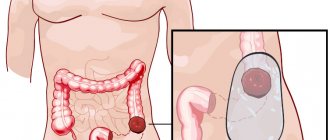
Malignant formations
Ileal cancer itself is not very common; metastasis from other organs is more often diagnosed. Nutrition plays an important factor in this disease.
It is fatty and smoked food, or rather its excessive consumption, that is a provoking factor. As mentioned above, if the disease is ignored for a long time, the tumor can spread and infect neighboring organs.
The following symptoms may appear:
- nausea;
- colic;
- bloating;
- heaviness;
- weight loss;
- obstruction;
- bleeding in the intestine.
Ulcer
In case of chronic intestinal disease and untimely, neglectful attitude to treatment, defects - ulcers - can form. According to statistics, more than 70% of the disease occurs in men.
https://www.youtube.com/watch?v=3ACyAyM-VoM
The main reasons may be disturbances in the functioning of blood vessels, taking medications that are not suitable, injuries to the intestinal lining, and excessive amounts of hydrochloric acid.
In the first stages, the characteristic symptoms are pain, which worsens when eating fatty and spicy foods, most often on the right side. In the final stages, pus and blood appear in the stool. If treatment is not started in time, complications may occur:
- perforation;
- cancer;
- narrowing of the intestine;
- internal bleeding.
Atresia
A characteristic symptom is that substances do not pass through the last part of the intestine. The peculiarity of this disease is that it is congenital.
Symptoms:
- bloating;
- lack of meconium;
- vomiting, nausea (typical already in the first days of life);
- anxiety;
- the child’s appetite disappears;
- insufficient feces.
Sections of the small intestine
The small intestine has 3 sections:
The initial section of the small intestine is the duodenum. It distinguishes between the upper, descending, horizontal and ascending parts. The small intestine and ileum do not have a clear boundary between themselves.
The beginning and end of the small intestine are attached to the posterior wall of the abdominal cavity. Throughout the rest of its length it is fixed by the mesentery. The mesentery of the small intestine is the part of the peritoneum that contains blood and lymphatic vessels and nerves and allows intestinal motility.
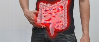
Pain in the right iliac region of the abdomen
Pain is the most striking symptom of any problem in the body. Thus, the body signals to the consciousness “Attention! A disaster has occurred! Save yourself! Unfortunately, abdominal pain is much more difficult to cope with than, for example, the sensation of a burned finger. Therefore, you should know what it can be, why it occurs and who to turn to for treatment.
What is pain in the lower abdomen? Where does it hurt? When did you get sick? Is this the first time such pain has occurred? Nature of pain in the lower abdomen Surgical causes of pain in the lower abdomen Appendicitis Terminal ileitis, or Crohn's disease Diverticulitis Ulcerative colitis Intestinal tumors Mesadenitis
Adhesive disease
What is pain in the lower abdomen?
To fully understand the situation, doctors will definitely ask several questions about pain:
- Where does it hurt?
- When did you get sick?
- Is this the first time such pain has occurred or has it happened before?
- How does the patient feel this pain?
In addition, it is important to find out the features of the increase in pain, the movement of the point of pain from place to place, additional symptoms that arise along with the pain syndrome, etc. The information received from an experienced specialist will certainly form a complete picture and additional diagnostic measures will only confirm the initial assumptions.
Where does it hurt?
Doctors conventionally divide the abdomen into three floors, each of which contains certain organs. Each floor is also divided into three areas. In the lower abdomen there are:
- right iliac;
- suprapubic;
- left iliac region.
In the right iliac region there is the cecum with the vermiform appendix extending from it, as well as the final part of the small intestine.
The left ileal region contains the sigmoid colon, as well as part of the jejunum and ileum.
The loops of the small intestine are also located above the pubis, but when the bladder is full it also protrudes into this area. Also, during pregnancy, the uterus emerges into the suprapubic region.
The lower part of the abdomen is limited in front by the rectus and oblique abdominal muscles, and in the back by the spine and muscles of the lumbar region.
Usually, if pain occurs, for example, above the pubis, then the organ that caused it is located in the same area. However, deviations from this rule are also possible.
When did you get sick?
For diagnosis, it is important to associate the onset of pain with some event in the patient’s life. For example, with some gynecological diseases, pain in the lower abdomen may occur after sexual intercourse.
In addition, knowledge of the moment of pain onset allows us to make assumptions about what is happening in the abdomen at the present time. So if a person suffered abdominal pain for 3-4 days, then the likelihood of appendicitis is extremely low - during this time the patient would either die or be in critical condition.
Is this the first time such pain has occurred?
If exactly the same pain sensations have occurred before, then there is a possibility that the process is chronic. This knowledge allows you to change treatment tactics and, often, avoid unnecessary surgery.
Nature of pain in the lower abdomen
The nature of the pain is perhaps the main criterion that any doctor clarifies in detail. The pain may be:
- constant;
- paroxysmal;
- aching;
- burning;
- pressing;
- piercing;
- light or, on the contrary, intense, etc.
By carefully analyzing the nature of the pain, you can make a fairly accurate assumption about its causes and prescribe adequate treatment.
Surgical causes of lower abdominal pain
There are a number of surgical diseases that lead to pain in the lower abdomen. Some of them are indications for immediate surgery, others can be treated conservatively. This is the most dangerous category of diseases, since at any moment they can lead to the development of life-threatening complications.
Appendicitis
This disease is the best known and most underestimated by “mere mortals”. Often people do not pay attention to mild pain in the right iliac fossa until the situation begins to seriously worsen.
With appendicitis, the pain is constant at first, often occurring in the upper abdomen or near the navel, simulating the symptoms of gastritis or intestinal colic.
After a few hours, the pain moves to the right iliac region, where it becomes burning, stabbing or pressing.
Coughing, straining, sudden movements and walking intensify them, forcing the patient to take a forced position - half-bent with the hand pressed to the sore spot. In bed, the patient always lies in the fetal position on the right side - this reduces pain.
Source: https://menchov.ru/bolit-pravaja-podvzdoshnaja-oblast-zhivota/
Lymphatic vessels
The lymphatic vessels of the small intestine begin in the villi of the mucous membrane; upon leaving the wall of the small intestine they enter the mesentery. In the mesenteric area, they form transport vessels that are capable of contracting and pumping lymph. The vessels contain a white liquid similar to milk. That's why they are called milky. At the root of the mesentery are the central lymph nodes.
Some lymphatic vessels may empty into the thoracic stream, bypassing the lymph nodes. This explains the possibility of rapid spread of toxins and microbes through the lymphatic route.
Transanal ileoscopy
Transanal ileoscopy is the traditional name for terminal ileoscopy, performed by inserting an endoscopic probe through the anus. To conduct this study, a special maternal colonoscope is used in conjunction with a daughter apparatus.
Transanal ileoscopy involves examining the lower ileum by retrogradely advancing the maternal endoscope through the valve of Bauhin. The maternal apparatus has a special biopsy channel through which, after passing through the ileocecal valve, a more flexible and mobile daughter “babyscope” is passed into the ileum.
This technique allows you to advance the probe to the required distance while maintaining maximum visualization of all parts of the ileum being examined.
However, it should be noted that the maternal probe has a rather rigid body and a large diameter (0.16 cm), therefore, in a number of diseases of the small intestine (Crohn's disease, polyposis, ulcerative colitis), its movement through the intestine is associated with certain difficulties. This is due to the presence of pathologically altered intestinal loops, their fusion, and narrowing of the loops due to adhesions and kinks. In such pathological conditions, transanal ileoscopy becomes technically impossible.
In such cases, the maternal endoscope is replaced with a transintestinal guide. The transintestinal guidewire has a much smaller diameter and is made of flexible, pliable Teflon. A small weight is attached to the distal end of the transintestinal tube, which makes it easier for the patient to swallow it.
The use of transintestinal intubation allows terminal ileoscopy to be performed according to indications directly in situations where it is not possible to perform transanal ileoscopy.
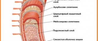
Mucous membrane
The mucous membrane of the small intestine is lined with single-layer prismatic epithelium.
Epithelial renewal occurs in different parts of the small intestine within 3-6 days.
The cavity of the small intestine is lined with villi and microvilli. Microvilli form the so-called brush border, which provides the protective function of the small intestine. Like a sieve, it sifts out high-molecular toxic substances and does not allow them to penetrate the blood supply and lymphatic system.
Symptoms
In the initial stage, ileitis can occur latently for some time, occasionally disturbing the patient with symptoms of a harmless intestinal disorder. Most often, people do not pay attention to such signs, but believe that this is normal overeating or a diet disorder.
Nevertheless, acute ileitis has quite clear symptoms:
- increased gas formation, accompanied by rumbling in the stomach;
- severe diarrhea, which can occur more than 10 times a day, is dangerous because it can cause dehydration;
- nausea and vomiting that does not bring relief;
- abdominal pain resembling appendicitis;
- increase in body temperature, often slight;
- increased fatigue;
- a sign of intoxication is headaches that do not stop after taking analgesics.

All of these symptoms are similar to many diseases of the digestive tract, so it is not possible to independently diagnose. If you have such signs, you should consult a doctor.
If the first symptoms are ignored, the disease becomes chronic, and the clinical picture is blurred:
- pain in the ileum becomes moderate, localized mainly in the navel area;
- diarrhea occurs rarely, but has a characteristic appearance - watery, with pieces of food that have not been digested;
- the patient loses weight for no apparent reason, this is due to impaired absorption of nutrients;
- Vitamin deficiency develops.
Symptoms can easily be confused with other pathologies of the digestive tract, and dehydration as a result of diarrhea can cause serious complications, so you need to contact a specialist for diagnosis and treatment.
Functions
The small intestine performs the most important functions for the body, such as
- digestion
- immune function
- endocrine function
- barrier function.
Digestion
It is in the small intestine that the processes of food digestion occur most intensively. In humans, the digestion process practically ends in the small intestine. In response to mechanical and chemical irritations, the intestinal glands secrete up to 2.5 liters of intestinal juice per day. Intestinal juice is secreted only in those parts of the intestine in which the food lump is located. It contains 22 digestive enzymes. The environment in the small intestine is close to neutral.
Fright, angry emotions, fear and severe pain can slow down the functioning of the digestive glands.
Food contains proteins, fats, carbohydrates and nucleic acids. For each component, there is a set of enzymes that can break down complex molecules into components that can be absorbed.
Absorption in the small intestine occurs throughout its entire length as food masses move through. Calcium, magnesium, and iron are absorbed in the duodenum; mainly glucose, thiamine, riboflabin, pyridoxine, folic acid, and vitamin C are absorbed in the jejunum. Fats and proteins are also absorbed in the jejunum.
Vitamin B12 and bile salts are absorbed into the ileal cavity. The absorption of amino acids is completed in the initial parts of the jejunum. Digestion in the human small intestine is the most important and at the same time the most complex function.
The immune system
It is difficult to overestimate the importance of the immune function of the intestines for maintaining the health of the body. It provides protection against food antigens, viruses, bacteria, toxins and drugs.
The mucous membrane of the small intestine contains more than 400 thousand per square meter. mm of plasma cells and about 1 million per square meter. see lymphocytes. This means that in addition to the epithelial layer that separates the external and internal environment of the body, there is also a powerful leukocyte layer.
Cells of the small intestine produce a number of immunoglobulins, which are absorbed on the mucous membrane and provide additional protection, forming the body's immunity.
Endocrine system
The small intestine is an important endocrine organ.
The number of endocrine cells in the small intestine is no less in mass than in such endocrine organs as the thyroid gland or adrenal glands.
More than 20 hormones and biologically active substances that control the functions of the gastrointestinal tract have been studied. In addition, it is known how they act in the body. The network of neurons located in the intestinal wall regulates intestinal functions with the help of various neurotransmitters, and is called the intestinal hormonal system.
Protective function
The process of breakdown of nutrients includes not only the supply of plastic and energy materials, but there is a danger of toxic substances entering the internal environment of the body. Foreign proteins pose a particular danger. During the process of evolution, a powerful protective system has formed in the gastrointestinal tract.
The effectiveness of the barrier function of the small intestine depends on its enzymatic activity, immune properties, the presence and condition of mucus, structural integrity, and degree of permeability.
When proteins are consumed as a result of breakdown, they lose their antigenic properties, turning into amino acids. But some of the proteins can reach the distal parts of the intestine. And here the level of permeability of the small intestine plays an important role. If permeability is increased, then the risk of penetration of antigens into the internal environment of the body increases.
The permeability of the intestinal wall increases with prolonged fasting, with inflammatory processes and especially with a violation of the integrity of the mucous membrane.
With limited penetration of food antigens, the body forms a local immune response, producing antibodies. Secretory antibodies form non-absorbable immune complexes with most antigens, which are then broken down into amino acids.
The permeability of the small intestine may increase with expanded intercellular space. This leads to hypersensitivity to food proteins, which is often a trigger for diseases such as allergies.
Proteins found in cereals, soybeans, and tomatoes have the ability to penetrate the intestinal barrier. They are extremely poorly broken down and have a toxic effect on the intestinal epithelium.
Normally, the barrier of the small and large intestines is almost completely insurmountable for microorganisms. But with poor nutrition, hypothermia, intestinal ischemia, and damage to the mucous membrane, a significant number of bacteria can overcome the intestinal barrier and enter the lymph nodes, liver, and spleen.
With a nutritional deficiency of essential amino acids and vitamin A, the normal renewal of the mucous membrane is disrupted.
In addition to its direct functions, the small intestine influences neighboring organs, regulating their activity. Through functional connections, it coordinates the interaction of all parts of the digestive system.
Diet menu for intestinal inflammation
Sample menu for loose stools:
Monday
- breakfast - chicken meatballs, buckwheat porridge, compote (cottage cheese for a snack).
- lunch - diet soup, fish soufflé, sweet tea (you can have a snack with tea and crackers).
- dinner – cottage cheese-buckwheat casserole, tea, crackers (jelly before bed).
Tuesday
- breakfast – boiled veal, stewed zucchini, compote (snack – dry biscuits, sweet tea)
- lunch - noodle soup, rice casserole with meat, tea (snack - jelly).
- dinner - stewed zucchini, stewed fish, compote (jelly before bed).
Wednesday
- cottage cheese, boiled rice, compote (snack – baked apples)
- lunch - soup with vegetables and barley, stewed fish, vermicelli, tea (snack - crackers, juice diluted with water)
- dinner – vegetable stew, boiled fish, tea (jelly before bedtime)
Thursday
- breakfast – mashed potatoes, chicken meatballs, apple compote (snack – berries)
- lunch – chicken noodles, vegetable stew, tea (snack – crackers, juice diluted with water).
- dinner - fish cutlets, stewed cauliflower, fruit soufflé, tea (jelly before bed).
Friday
- breakfast – grated cottage cheese, semolina porridge, compote (snack – baked apple)
- lunch – vegetable soup, meatloaf stuffed with steamed omelet, tea (snack – tea, crackers)
- dinner - semolina casserole, fish balls, compote (jelly before bed).
Diagnosis and treatment
It is difficult to diagnose ileitis based on symptoms; a physical examination is necessary, which in itself carries little information, but is mandatory:
- it is necessary to pay attention to the condition of the patient’s tongue - a dry organ with a white coating is a sign of inflammation of the digestive organs;
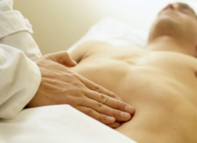
- upon palpation, the patient feels pain in the iliac region on the right;
- if you tap a swollen belly, the sound will be ringing;
- You can hear the stomach rumbling, especially in the navel area.
The structure of the stool will be liquid, light yellow in color, with pieces of food. Instrumental research has difficulties due to the location of the organ, and therefore is not informative, but is used to differentiate the disease from other pathologies.
A laboratory blood test confirms the presence of an inflammatory process in the body, and a stool test can identify the causative agent of ileitis; if the stool contains hidden blood, this indicates internal bleeding.
Diagnostics
If ileitis is suspected, the patient undergoes a laboratory examination, including:
- general blood test (leukocytosis with a shift in the leukocyte formula to the left, increased ESR);
- bacteriological and virological examination of feces - allows you to identify the causative agent of the disease and determine its sensitivity to antibacterial drugs;
- coprogram (decreased enzymatic activity, large amounts of carbohydrates and undigested muscle fibers);
- feces for occult blood;
- biochemical blood test (hypoproteinemia, decreased concentration of some microelements).
If ileitis is suspected, an X-ray examination of the intestine with a contrast agent (barium sulfate) is indicated. By assessing the characteristics of the passage of barium suspension through the intestines, spasmodic areas of the ileum, the presence of strictures and fistulas are revealed.
In 70% of cases, regular pain in the right iliac region is caused by chronic ileitis, most often of yersinia etiology.
Considering that ileitis in many cases is accompanied by other diseases of the gastrointestinal tract, FEGDS, ultrasound and multispiral computed tomography of the abdominal organs are indicated.
Ileitis is differentiated from other pathologies that also occur with diarrhea:
Causes
With inflammation of the ileum, the disease in children usually has an acute course and is provoked by pathogenic microorganisms (streptococcus, staphylococcus) or viruses. But in adults, ileitis mainly occurs in a chronic form with temporary exacerbations. Yersinias often become provocateurs. And quite rarely it can be caused by parasites.
The expected factors for the development of ileitis are:
- allergy;
- unhealthy diet (eating fatty, fried, spicy foods);
- bad habits (smoking, alcohol);
- associated pathologies of the digestive tract (duodenitis, pancreatitis);
- various operations on the intestines;
- fermentopathy;
- gastrointestinal infections;
- inactive, sedentary lifestyle (working at a computer);
- intoxication of the body with chemicals.
The cause of ileitis may be the presence of typhoid fever, ulcerative colitis, Crohn's pathology (leading to terminal ileitis), or tuberculosis. In this situation, ileitis is considered a symptom of the underlying pathology.
Long-term chronic ileitis leads to severe malabsorption of nutrients. This turns out to be the cause of hypovitaminosis and osteoporosis. It also leads to brittle hair, nails, and weight loss.